Most cardiac arrest algorithms are built around the heart. We think ventricular fibrillation, we think pulseless VT, we think defibrillation and epinephrine. But a significant subset of cardiac arrests — particularly in trauma bays, post-operative suites, and labor-and-delivery units — are not fundamentally electrical problems at all. They are plumbing failures. The heart has stopped because there is nothing left to pump.
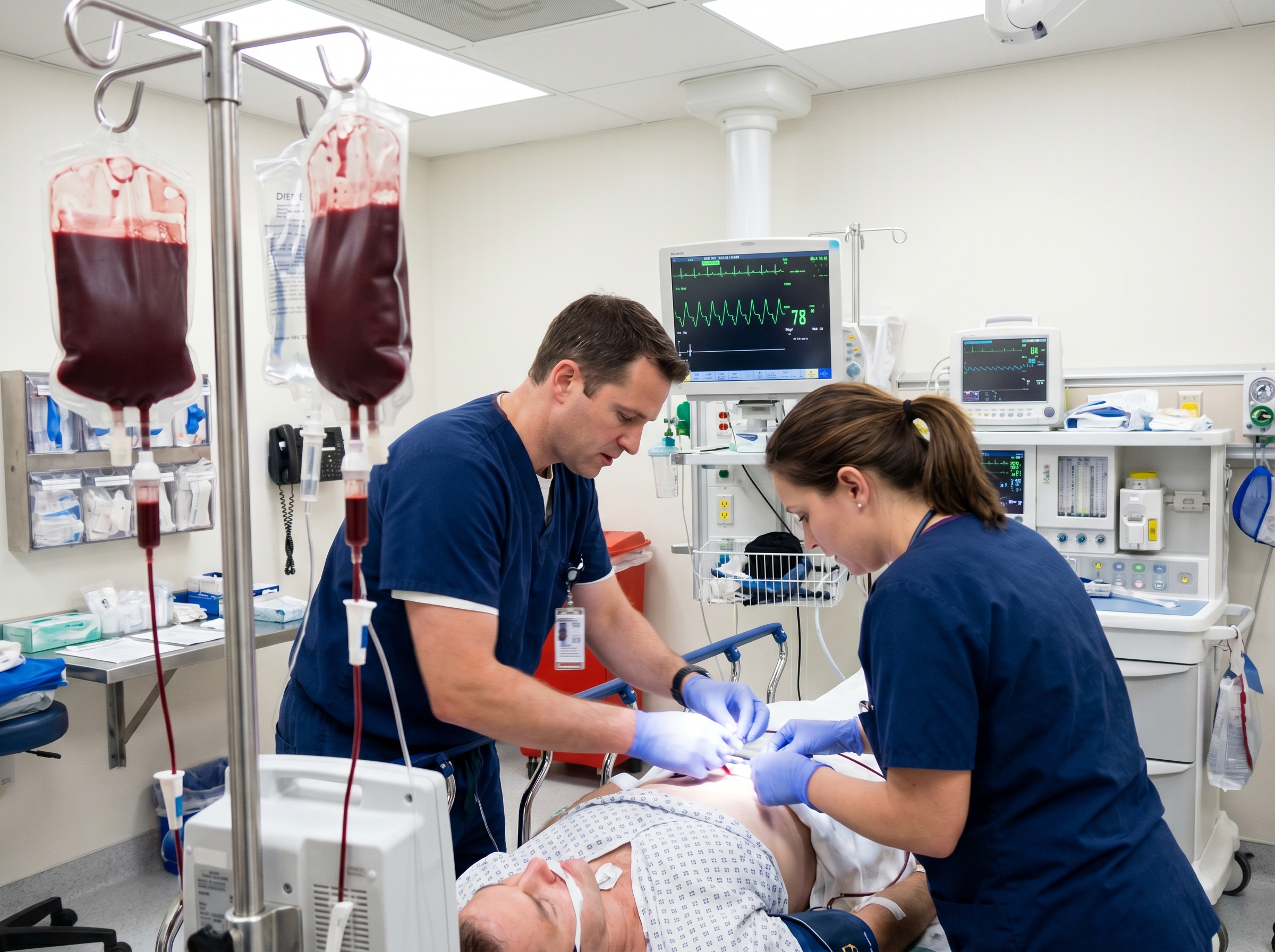
Hemorrhagic shock-driven cardiac arrest represents one of the most challenging and time-critical resuscitation scenarios in emergency medicine. When hypovolemia from catastrophic blood loss is the precipitating cause of pulseless electrical activity or asystole, standard ACLS algorithms are necessary but profoundly insufficient on their own. You can push epinephrine every three to five minutes and run perfect high-quality CPR, but if the patient's intravascular volume is critically depleted and coagulopathy is consuming whatever clot-forming capacity remains, you are fighting the pathophysiology with one hand tied behind your back.
This article is written for the emergency physicians, intensivists, trauma surgeons, and advanced practice providers who find themselves at the center of these codes — the clinicians who need to run a simultaneous ACLS algorithm AND a massive transfusion protocol, manage a team that spans the trauma bay and the blood bank, and make rapid decisions under extreme uncertainty. We will cover the physiology of hemorrhagic cardiac arrest, how massive transfusion protocols integrate with ACLS, the evidence base driving modern balanced resuscitation, and the team dynamics that determine survival.
To manage hemorrhagic cardiac arrest effectively, you have to understand what is actually happening at the cellular and hemodynamic level. Hemorrhagic shock exists on a spectrum. Class I and II shock are compensated states where the body can maintain perfusion through tachycardia and vasoconstriction. Class III and IV shock — representing losses of 30 to 40 percent or greater than 40 percent of circulating blood volume respectively — overwhelm compensatory mechanisms. Cardiac output collapses, coronary perfusion pressure drops, and the myocardium itself becomes ischemic.
Simultaneously, the trauma-induced coagulopathy (TIC) that accompanies major hemorrhage creates a vicious cycle. Acidosis, hypothermia, and dilution — the so-called "lethal triad" — impair clotting factor function and platelet aggregation at the precise moment when effective coagulation is most critical. The result is ongoing hemorrhage from a patient who can no longer form effective clots, compounding the hypovolemia that is driving the arrest. This is why the reversible causes of cardiac arrest — the Hs and Ts — place hypovolemia at the top of the list. In the hemorrhagic arrest patient, H for hypovolemia is not just one consideration among eight; it is the diagnosis, and every other intervention is subordinate to addressing it.
Pulseless electrical activity in the setting of hemorrhage is particularly instructive. The heart retains electrical organization — sometimes a near-normal sinus rhythm on the monitor — but mechanical function has failed because the ventricles have insufficient preload to generate forward flow. Understanding PEA and its reversible causes is essential here: in the trauma patient with PEA, an organized rhythm on the monitor should immediately prompt you to think hemorrhage, tension pneumothorax, and tamponade — in that order of probability — before you reach for the epinephrine.
A massive transfusion protocol (MTP) is a pre-activated, institutionally standardized system for the rapid delivery of large volumes of blood products in a balanced ratio. The traditional definition of massive transfusion is the administration of ten or more units of packed red blood cells within twenty-four hours, but in clinical practice, activation criteria are trigger-based and prospective — you cannot wait for a retrospective tally before you start delivering products.
The ACS TQIP Massive Transfusion in Trauma Guidelines recommend that every trauma center develop a multidisciplinary MTP with clearly defined activation triggers, a protocol for the preparation and delivery of blood products in a predetermined ratio, and a system for ongoing monitoring and deactivation. The Assessment of Blood Consumption (ABC) score is a widely validated activation tool: a pulse greater than 120 bpm, systolic blood pressure below 90 mmHg, a positive FAST examination, and penetrating torso injury each earn one point, with a score of two or more indicating high probability of requiring massive transfusion. In the arrested patient, of course, you are already beyond scoring — MTP activation should be simultaneous with resuscitation initiation.
Products typically arrive from the blood bank in pre-packaged coolers or sequential rounds containing predetermined ratios of packed red blood cells (PRBCs), fresh frozen plasma (FFP), and platelets. The speed of delivery is as important as the composition: the blood bank must understand that MTP activation means continuous product preparation without waiting for individual orders.
The modern approach to massive transfusion has been shaped decisively by two landmark trials: PROMMTT and PROPPR. The PROPPR randomized clinical trial, published in JAMA, enrolled 680 severely injured patients at twelve Level I trauma centers across North America. Patients were randomized to receive plasma, platelets, and red blood cells in either a 1:1:1 ratio or a 1:1:2 ratio — that is, one unit of plasma and one apheresis platelet unit per unit of PRBCs versus a PRBC-heavy approach.
The primary mortality endpoints — death at 24 hours and at 30 days — did not reach statistical significance between groups (12.7% vs. 17.0% at 24 hours; 22.4% vs. 26.1% at 30 days). But the critical secondary finding was unmistakable: fewer patients in the 1:1:1 group died from exsanguination by 24 hours (9.2% vs. 14.6%, p=0.03), and more patients achieved hemostasis. The trial established that a balanced 1:1:1 resuscitation strategy reduces death from ongoing hemorrhage — the precise mechanism that drives hemorrhagic cardiac arrest — even when the overall mortality curves converge at 30 days.
The Eastern Association for the Surgery of Trauma (EAST) Practice Management Guidelines for damage control resuscitation synthesize this evidence into actionable recommendations. Key guidance includes: implementing a massive transfusion or damage control resuscitation protocol at trauma centers; targeting high plasma-to-RBC and platelet-to-RBC ratios during empiric early resuscitation; limiting crystalloid administration; and considering permissive hypotension in penetrating trauma patients without traumatic brain injury. The EAST guidelines also conditionally recommend tranexamic acid (TXA) when administered within three hours of injury — a window that can close rapidly once a patient progresses to cardiac arrest.
Here is where the clinical complexity becomes acute. A patient in hemorrhagic cardiac arrest requires you to run two complex, parallel protocols simultaneously — ACLS and MTP — while managing a team that spans the bedside and the blood bank. Neither protocol can be subordinated to the other. High-quality chest compressions maintain coronary perfusion pressure and buy time. But if you achieve ROSC without volume, the heart will immediately re-arrest into a PEA or asystolic rhythm because there is still nothing to pump.
The organizational principle that works in practice is explicit role assignment. The team leader calls the code and owns the ACLS algorithm. A second physician or senior provider owns the MTP — they are on the phone with the blood bank, tracking product delivery, monitoring the ratio, and managing adjuncts including TXA, calcium, and vasopressors. Clear ACLS team communication scripts are the infrastructure that allows this dual-track resuscitation to function without confusion or crossed communications at a moment when every second counts.
Practically speaking, the integration of MTP and ACLS during active cardiac arrest from hemorrhage involves these parallel tracks:
The 1:1:1 ratio of PRBCs:FFP:platelets represents the current standard of care for empiric hemostatic resuscitation based on PROPPR and the ACS TQIP guidelines. In practice, this means that for every unit of PRBCs delivered, a unit of FFP and an apheresis platelet unit (or equivalent pooled platelets) should accompany it. The goal is to replicate whole blood as closely as possible — which is why there is growing institutional interest in low-titer group O whole blood (LTOWB) as a streamlined alternative that inherently delivers a balanced product without the logistical overhead of ratio management.

Calcium is a frequently overlooked but critically important adjunct in massive transfusion. Citrate, used as a preservative in stored blood products, chelates ionized calcium. With rapid, high-volume transfusion, citrate load can outpace hepatic metabolism and produce clinically significant hypocalcemia — which impairs myocardial contractility and coagulation factor function simultaneously. Empiric calcium chloride (1g IV) or calcium gluconate should be administered with each round of MTP products. If arterial access is in place, trending ionized calcium directly is the preferred approach in arrested or peri-arrest patients.
Avoid the crystalloid trap at all costs. Historical resuscitation practices relied heavily on normal saline and lactated Ringer's. While a modest crystalloid bolus may bridge the gap before blood products arrive, large-volume crystalloid resuscitation in the hemorrhagic arrest patient is actively harmful. Crystalloids dilute clotting factors, worsen acidosis through hyperchloremic mechanisms, and contribute directly to the lethal triad of coagulopathy, acidosis, and hypothermia. The StatPearls review of massive transfusion summarizes the evidence clearly: crystalloid-heavy resuscitation increases coagulopathy and mortality in the hemorrhagic shock patient. Get blood products moving, and minimize crystalloid to the minimum necessary bridge.
Hemorrhagic cardiac arrest does not occur exclusively in the trauma bay. Several distinct clinical contexts require modified thinking and adapted protocol approaches, each with specific physiologic considerations that alter standard MTP management.
Obstetric Hemorrhage: Postpartum hemorrhage (PPH) is one of the leading causes of maternal mortality worldwide, and severe PPH can progress to cardiac arrest with devastating speed. Maternal cardiac arrest protocols must account for aortocaval compression by the gravid uterus, the need for left lateral uterine displacement during CPR, and the unique physiologic changes of pregnancy that alter normal vital sign thresholds. MTPs in obstetric settings should be specifically calibrated for this population, with particular attention to fibrinogen levels — obstetric coagulopathy often features disproportionate hypofibrinogenemia requiring cryoprecipitate administration in addition to standard MTP product rounds.
Gastrointestinal and Aortic Hemorrhage: Massive upper or lower GI hemorrhage and ruptured aortic aneurysms are non-trauma causes of hemorrhagic cardiac arrest encountered in emergency departments with regularity. These patients may present without the dramatic mechanism that triggers trauma activation, leading to delayed recognition and late MTP activation. A high index of suspicion in hypotensive, tachycardic patients with known aneurysmal disease or active GI bleeding is essential. In many of these cases, the arrest is foreseeable and preventable with earlier, more aggressive intervention.
Burn Patients with Concurrent Trauma: Massive burns with concurrent traumatic injury represent another modified resuscitation context. Burn unit ACLS protocols are distinct from standard trauma resuscitation and require familiarity with the Parkland formula, the risks of fluid creep, and the unique airway management challenges of thermal and inhalation injury superimposed on hemorrhagic physiology.
Point-of-care testing transforms MTP management from empiric to goal-directed as rapidly as the laboratory turnaround allows. Thromboelastography (TEG) and rotational thromboelastometry (ROTEM) provide real-time assessment of clot formation, clot strength, and fibrinolysis that cannot be derived from standard coagulation panels. These viscoelastic tests allow the resuscitation team to identify specific deficits — is this primarily a clotting factor deficiency, a platelet function problem, or fibrinolytic activation? — and tailor product delivery accordingly rather than continuing empiric ratio transfusion beyond what the specific coagulopathy demands. Arterial blood gas interpretation during active resuscitation provides critical real-time data on lactate, pH, base deficit, and ionized calcium — all of which guide ongoing management and provide prognostic context.
Lactate clearance is one of the most reliable markers of resuscitation adequacy. A lactate trending toward normal indicates that tissue perfusion is being restored. A persistently elevated or rising lactate during active transfusion suggests either ongoing hemorrhage outpacing replacement volume, inadequate product delivery rate, or a source that has not been controlled. Serial lactate measurements every 30 to 60 minutes during active resuscitation are a minimum standard; more frequent sampling via arterial line is preferable in arrested or peri-arrest patients.
When ROSC is achieved in the hemorrhagic arrest patient, the work is far from over. The post-ROSC patient who arrested from hemorrhage is at extreme risk for re-arrest from continued bleeding, coagulopathy, myocardial stunning, and secondary inflammatory effects of ischemia-reperfusion injury. Maintaining adequate MAP — typically 65 mmHg or higher, adjusted downward if permissive hypotension remains appropriate for the injury pattern — while continuing balanced blood product delivery, pursuing definitive hemorrhage control in the operating room or interventional suite, and monitoring for complications of massive transfusion including TRALI, TACO, and citrate toxicity are the simultaneous priorities of the immediate post-arrest phase.
MTP documentation in the setting of cardiac arrest demands the same rigor as any high-stakes resuscitation — and the complexity of running two simultaneous protocols raises the documentation burden considerably. Times of MTP activation, product delivery rounds, total volumes administered by product type, adjunct medication administration including TXA and calcium, laboratory values, and times to ROSC or termination of resuscitation must all be captured contemporaneously by a designated recorder. ACLS documentation best practices apply with full force in the MTP context: real-time, role-assigned documentation is non-negotiable, because retrospective reconstruction of a dual-protocol resuscitation is both inaccurate and legally inadequate.
MTP deactivation is as important as activation. Continued unnecessary blood product delivery after hemorrhage is controlled exposes patients to transfusion-associated complications without clinical benefit. Deactivation criteria should be pre-specified in the institutional protocol and typically include: hemostasis clinically achieved, hemoglobin stabilized at an acceptable threshold, coagulation parameters normalized on TEG/ROTEM or standard labs, and surgical source control confirmed. The blood bank should receive explicit deactivation notification to stop preparing further product rounds, with tracking of total products issued for quality reporting.
Every MTP activation — whether it ends in survival, death, or termination of resuscitation — should undergo multidisciplinary review. A comprehensive review of massive transfusion and major hemorrhage protocols in ScienceDirect emphasizes that systematic protocol refinement through case review is one of the primary mechanisms by which institutions improve MTP outcomes over time. Time from arrest recognition to first blood product delivery, adherence to ratio targets, accuracy of activation criteria, and communication failures between clinical teams and the blood bank are all productive targets for quality improvement cycles.
The dual complexity of running simultaneous ACLS and MTP algorithms under crisis conditions demands a team that has deeply internalized both protocols. The behavioral automaticity required to manage airway, compressions, rhythm checks, epinephrine intervals, MTP product tracking, calcium dosing, and surgical consultation simultaneously cannot be acquired by reading a policy document. It is built through deliberate training, simulation, and the kind of role-based team practice that structured ACLS education provides — practice where the cognitive load of executing the algorithm is reduced enough that the team leader can think about the pathophysiology, not just the protocol steps.
At Affordable ACLS, our certification course is developed and taught by board-certified emergency medicine physicians who manage these exact scenarios clinically. The curriculum covers the reversible causes of cardiac arrest — including hypovolemia — with the depth and clinical contextualization that allows providers to recognize hemorrhagic arrest rapidly and pivot their management accordingly. Our courses are fully AHA/ILCOR-aligned, available online for provider convenience, and priced to be accessible: ACLS at $99, BLS at $59, PALS at $99, with group pricing available for institutional teams. Reach us at 866-655-2157 to discuss training needs or institutional partnerships.
Hemorrhagic cardiac arrest is a resuscitation scenario that demands you hold two complex algorithms in your mind simultaneously, build a team capable of executing both without confusion, and recognize that standard ACLS interventions — while necessary — are categorically insufficient if the underlying hypovolemia and coagulopathy are not aggressively addressed in parallel. The rhythm on the monitor is almost a distraction from the real problem, which is happening in the vessels and the coagulation cascade, not the conduction system.
The evidence base is clear and consistent. Balanced resuscitation with a 1:1:1 ratio of PRBCs, FFP, and platelets reduces death from exsanguination. Early MTP activation — ideally before cardiac arrest, when the clinical trajectory is recognized — saves more lives than reactive activation after the patient has arrested. Tranexamic acid given within three hours preserves the coagulation system at its most vulnerable. Crystalloid restriction prevents the dilutional coagulopathy that compounds hemorrhagic shock into an irreversible state. And source control, whether surgical, interventional, or procedural, is the definitive treatment that allows everything else to succeed.
The next time you walk into a trauma bay or an ED resuscitation room and see an organized rhythm on the monitor with no pulse — and the mechanism is hemorrhage — remember that you are not fighting an electrical problem. You are fighting an empty tank. The solution is already on its way from the blood bank. Know your MTP. Know your team roles. Know your reversible causes. And train until the protocol becomes reflex. That is what your patient needs when hemorrhagic shock is the force driving the code.
.jpg)


