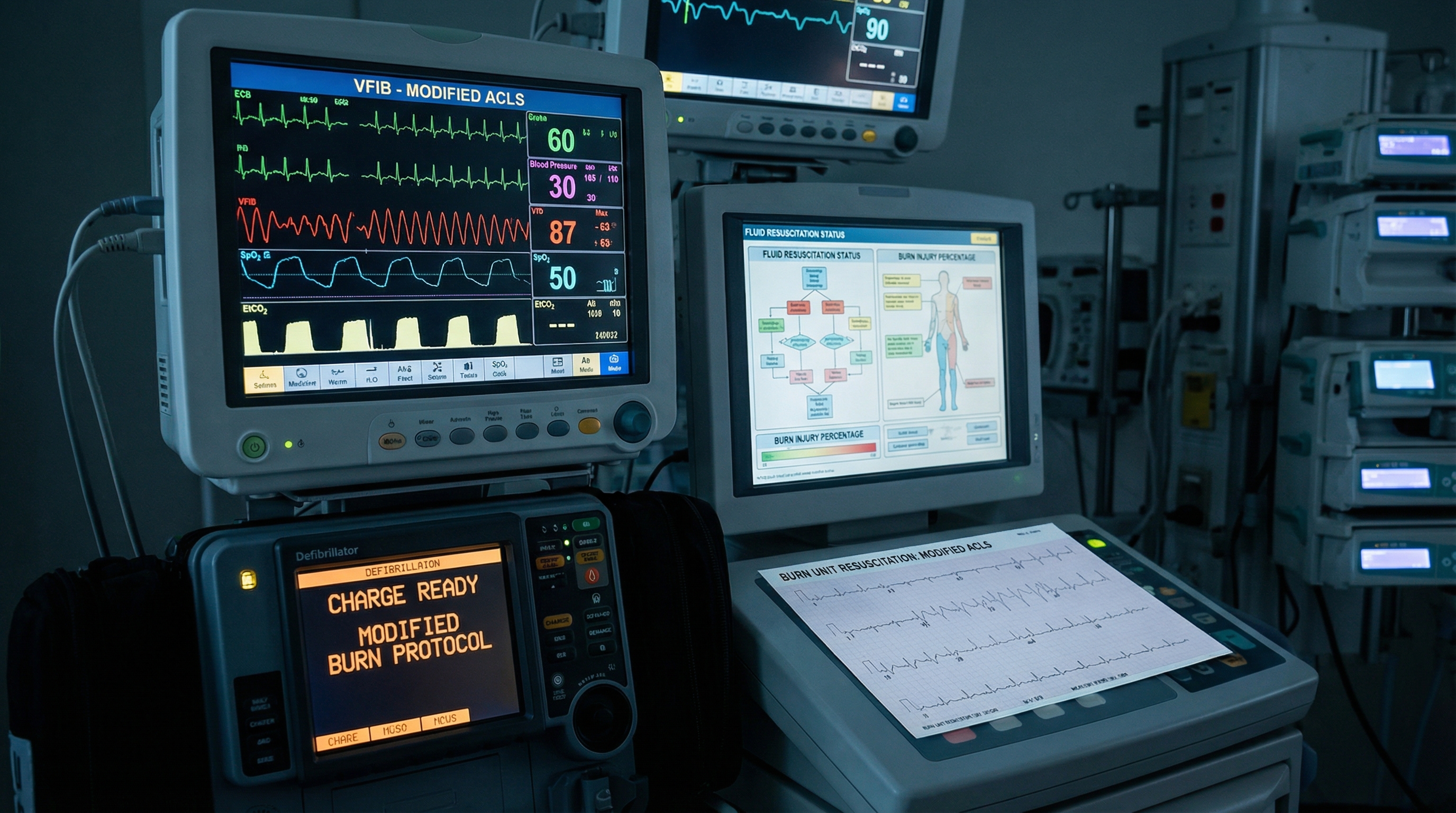
When a burn patient arrests, you are not running a standard ACLS code. You are navigating one of the most physiologically complex resuscitation scenarios in all of emergency medicine. The burned patient arrives with disrupted fluid balance, potential inhalation injury, electrolyte derangements, and a hyperinflammatory state that fundamentally alters how cardiac arrest develops and how it must be managed. Applying standard ACLS cardiac arrest algorithms without modification to this population is not just suboptimal — it can be dangerous.
For nurses, physicians, paramedics, and other advanced providers working in burn units, trauma centers, or emergency departments, a working knowledge of how standard protocols must shift in the burned patient is essential clinical currency. This article walks through the pathophysiology driving those differences, the specific modifications required at each step of resuscitation, and the post-arrest care priorities that separate survivors from non-survivors in this uniquely challenging patient population.
Cardiac arrest in burn-injured patients is poorly characterized compared to other special resuscitation circumstances, and the outcomes data is sobering. A 2025 scoping review published in the Scandinavian Journal of Trauma, Resuscitation and Emergency Medicine examined cardiac arrest following burn injury and found that outcomes remain uniformly poor in most burn etiologies — with the notable exception of electrical injury, where meaningful survival is possible with prompt resuscitation.
Burn-associated cardiac arrest can occur in several distinct phases. In the immediate phase (within the first hours), causes include direct electrical injury, explosion-related trauma, and severe hypoxia from inhalation injury. In the early resuscitation phase (first 24-72 hours), massive fluid shifts, hyperkalemia, and myocardial depression dominate the picture. Later in the hospital course, sepsis-driven cardiac dysfunction, abrupt electrolyte swings during wound debridement, and the cumulative cardiovascular toll of repeated surgical procedures create ongoing arrest risk. Each phase has its own dominant pathophysiology and demands a tailored response.
To modify a protocol intelligently, you first have to understand what is different inside the burned patient's cardiovascular system. Several interacting mechanisms make the burned myocardium uniquely vulnerable.
Myocardial depression is present in virtually all major burns. Research examining cardiac physiology changes after severe burn injury describes slowed isovolemic relaxation, impaired contractility, and decreased diastolic compliance manifesting as reduced cardiac output with compensatory tachycardia and elevated peripheral vascular resistance. This dysfunction is not simply volume depletion — it reflects direct myocardial toxicity from inflammatory mediators, catecholamine surge, and cytokine release.
Hyperkalemia is arguably the most dangerous electrolyte threat in burn resuscitation. Massive tissue destruction releases intracellular potassium directly into the circulation. Prolonged immobility, rhabdomyolysis, and renal impairment compound this release. Elevated potassium destabilizes cardiac membranes, widening the QRS, flattening P waves, and ultimately precipitating ventricular fibrillation or pulseless electrical activity. Understanding the relationship between hyperkalemia and cardiac arrest is critical — our detailed guide on hyperkalemia-induced cardiac arrest recognition and emergency treatment covers how to identify and address this threat at every step of the resuscitation.
Inhalation injury is present in approximately 20-30% of hospitalized burn patients and dramatically worsens prognosis. Carbon monoxide poisoning causes tissue hypoxia by displacing oxygen from hemoglobin. Cyanide toxicity, particularly from smoke containing synthetic materials, disrupts cellular respiration at the mitochondrial level. Upper airway edema can progress from subtle to complete obstruction within hours of injury as the inflammatory cascade amplifies capillary leak in airway mucosa. Any of these mechanisms can precipitate or complicate cardiac arrest in ways that require specific countermeasures.
Massive fluid shifts occurring in the first 24-48 hours create a dynamic hemodynamic picture. The Parkland formula and its variants guide initial fluid resuscitation, but the transition between the first and second day — when capillary integrity begins to recover and administered fluid begins to return to the intravascular space — creates its own hemodynamic risks. Pulmonary edema, abrupt electrolyte shifts, and transiently elevated cardiac preload can all destabilize the patient during this phase. Comprehensive reviews of cardiovascular dysfunction in burns confirm that the cardiac complications of major burns are multifactorial and can persist well beyond initial resuscitation.
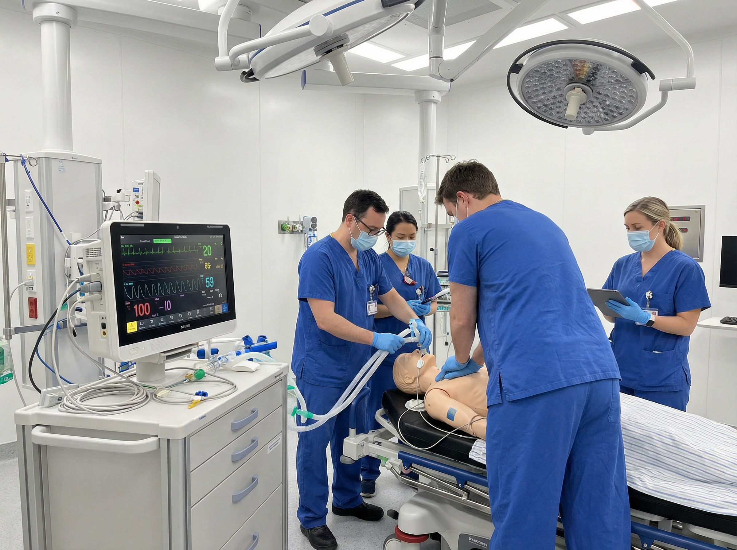
In standard ACLS, advanced airway placement is balanced against minimizing interruptions to chest compressions. In the burned patient — particularly one with suspected inhalation injury — early definitive airway management takes on an urgency that supersedes this typical calculus. Edema can progress with terrifying speed in the burn patient's airway. A patient who appeared to have a manageable airway on arrival may become impossible to intubate or even bag-mask ventilate within hours.
For providers managing a burn patient who has arrested or is peri-arrest, early direct laryngoscopy or video laryngoscopy is strongly preferred over prolonged bag-mask attempts when there is any sign of airway compromise — singed nasal hairs, stridor, carbonaceous sputum, hoarseness, or facial burns. Our comprehensive resource on mastering endotracheal intubation covers the technical aspects of securing a difficult airway under these high-pressure conditions.
Critical medication alert: succinylcholine is absolutely contraindicated in burn patients beyond the first 24 hours of injury. The mechanism is well-established and potentially fatal. Burn injury causes upregulation of extrajunctional nicotinic acetylcholine receptors throughout skeletal muscle. When succinylcholine binds these proliferated receptors, it triggers a massive, uncontrolled efflux of intracellular potassium into the bloodstream. As detailed in StatPearls' succinylcholine reference, in a patient already at risk for hyperkalemia from tissue destruction, this pharmacologically-induced potassium surge can precipitate ventricular fibrillation or cardiac standstill within minutes. This risk begins at 24 hours post-injury, peaks at 7-10 days, and — critically — may persist for years after the original burn. Rocuronium is the preferred neuromuscular blocking agent for rapid sequence intubation in burn patients, with sugammadex available for reversal if needed.
Every ACLS provider learns to systematically work through the reversible causes of cardiac arrest — the Hs and Ts. In burn patients, this differential takes on a different weighting and includes some burn-specific additions that must be front of mind. Understanding the full Hs and Ts framework is the foundation on which burn-specific modifications are built.
In burn patients, the following causes deserve elevated priority in your differential:
Standard high-quality CPR remains the foundation of burn resuscitation, but several practical challenges require adaptation. Burned chest wall tissue may have altered compliance, particularly in circumferential full-thickness chest burns with eschar formation. Eschar is rigid and inelastic; if it has not been addressed prior to arrest, effective chest compressions may be impossible. Emergency escharotomy of the anterior chest — longitudinal incisions through the eschar parallel to the sternum — may be necessary to allow adequate compression depth.
Pad placement for defibrillation presents its own challenge. Standard anterolateral or anteroposterior pad placement may not be feasible if those skin surfaces are burned. Providers should use the largest available unburned surface area on the chest for pad placement, accepting altered positioning to deliver effective shocks. The principle of high-performance CPR team coordination becomes especially important when the team must simultaneously manage airway edema, difficult IV access, escharotomy, and defibrillation — all while maintaining compression quality.
Vascular access in burn patients deserves special mention. Peripheral IV access through burned tissue is generally discouraged, but intraosseous access — tibial or humeral — is an excellent alternative when peripheral access is impossible. Central venous access, if time permits, provides reliable high-flow access for fluid and medication delivery during prolonged resuscitation.
The standard ACLS medication sequence — epinephrine, amiodarone for shockable rhythms — applies in burn patients with some important contextual modifications. Our ACLS medications reference guide covers standard dosing; what follows are the burn-specific overlays.
Epinephrine: Standard 1 mg IV/IO every 3-5 minutes as per ACLS guidelines. Note that burn patients often already have massively elevated endogenous catecholamine levels as part of the stress response to injury, which may blunt the expected pharmacological response. This does not change the dosing recommendation but contextualizes why some burn arrest patients are refractory.
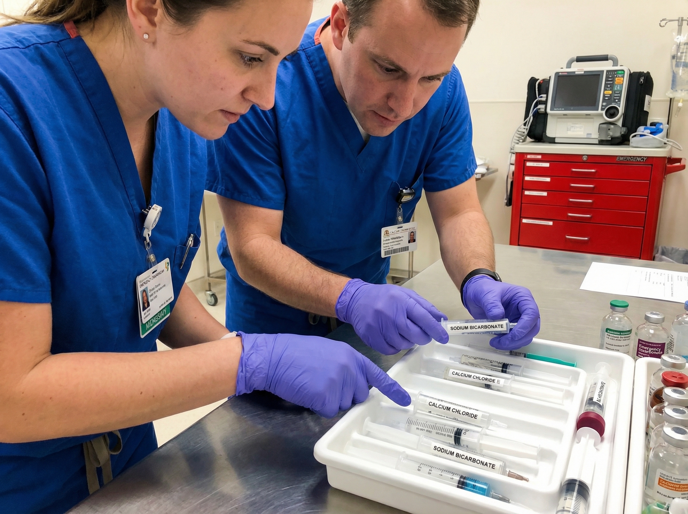
Calcium chloride or calcium gluconate: Should be added early in the medication sequence when hyperkalemia is suspected — which should be the default assumption in a burn arrest. Calcium stabilizes the myocardial membrane against the dysrhythmogenic effects of potassium, buying time while other interventions work. Calcium chloride 1g IV/IO provides three times the elemental calcium of an equivalent volume of calcium gluconate and is preferred in cardiac arrest situations.
Sodium bicarbonate: Has a role in the burn arrest that goes beyond the standard ACLS approach. In addition to treating hyperkalemia by shifting potassium intracellularly, bicarbonate addresses the profound metabolic acidosis common in burn patients and may improve responsiveness to catecholamines in an acidotic myocardium. One to two mEq/kg IV/IO is a reasonable empirical dose.
Hydroxocobalamin: For arrests with suspected cyanide toxicity, hydroxocobalamin 5g IV is the treatment of choice. It binds cyanide to form cyanocobalamin, which is renally excreted. Critically, unlike older cyanide antidotes (sodium nitrite, sodium thiosulfate), hydroxocobalamin does not cause methemoglobinemia and can be safely given when CO toxicity is also possible — making it ideal for the burn arrest setting where both toxins may be present simultaneously.
Amiodarone: Standard dosing applies for shockable rhythms. However, it is worth recognizing that ventricular fibrillation in the burn patient may be driven by hyperkalemia rather than a primary cardiac dysrhythmia. In this scenario, treating the underlying hyperkalemia is essential alongside antidysrhythmic therapy. Standard amiodarone dosing at 300 mg IV/IO for the first dose and 150 mg for subsequent doses remains the approach for refractory VF/VT, consistent with current 2025 AHA Adult Advanced Life Support Guidelines.
Electrical injury deserves specific mention because it represents the burn arrest scenario where aggressive resuscitation is most likely to result in meaningful survival. Unlike thermal burns, where cardiac arrest often reflects the end stage of overwhelming physiological insult, electrical-injury cardiac arrest frequently occurs in previously healthy individuals whose hearts have been thrown into ventricular fibrillation by the passage of current. The myocardium itself may be fundamentally intact.
Early defibrillation is the highest priority in electrical-injury VF. Prolonged resuscitation efforts are more justified than in most other burn arrest scenarios — cardiac arrest from electrical injury has been described as the one burn context where extended CPR with eventual ROSC and neurologically intact survival is a realistic goal. Do not terminate resuscitation efforts prematurely in electrical arrest. The standard ACLS principle of treating shockable rhythms with immediate defibrillation applies directly here.
Rhabdomyolysis is universal in significant electrical injury. Massive myoglobin release threatens renal function and can contribute to hyperkalemia. Aggressive hydration targeting urine output of 1-2 mL/kg/hour and urinary alkalinization to prevent myoglobin precipitation in renal tubules are standard post-arrest priorities in this subtype.
Achieving return of spontaneous circulation is only the beginning. Post-ROSC care in the general population focuses on targeted temperature management, hemodynamic optimization, and neuroprotection. In burn patients, all of these objectives are present but require burn-specific modification.
Targeted temperature management (TTM) after cardiac arrest in the general population involves cooling to 32-36°C. In burn patients, this approach is complicated by the fact that burn victims already lose body heat at an accelerating rate through damaged skin. Hypothermia is a known complication of burn resuscitation itself, and active rewarming — not cooling — is often necessary. The neuroprotective benefits of TTM must be weighed against the physiological impossibility of achieving and maintaining target temperatures in a patient losing heat through large surface area burns. Institution-specific protocols vary, and shared decision-making between the resuscitation team and burn specialists is essential.
Hemodynamic targets after ROSC in burn patients generally aim for mean arterial pressure greater than 65 mmHg, with awareness that volume requirements are typically larger than in non-burn patients. The myocardial depression described earlier means that vasoactive support with norepinephrine or vasopressin may be needed even after adequate volume replacement. Echocardiography, when available, guides the balance between volume resuscitation and vasoactive support.
Continuous monitoring of potassium, lactate, pH, and coagulation parameters is mandatory in the post-arrest burn patient. Hyperkalemia recurrence is common. Coagulopathy — driven by massive transfusion requirements, hypothermia, and acidosis — adds hemorrhagic risk during any required surgical intervention.
Burn-associated cardiac arrest has notoriously poor outcomes except in the specific scenarios described — primarily electrical injury and cases where a fully reversible cause (tension pneumothorax, tamponade, CO toxicity) is rapidly identified and corrected. The 2025 AHA guidelines acknowledge that special circumstances of resuscitation require individualized assessment of expected benefit.
Factors suggesting an extremely poor prognosis in burn cardiac arrest include: massive thermal burns with greater than 70-80% total body surface area (TBSA) involvement, prolonged resuscitation without ROSC, non-shockable initial rhythm (asystole or PEA) in thermal burns without an identified reversible cause, and the absence of electrical injury as an etiology. These factors should inform — but not automatically determine — decisions about continuation or termination of efforts. Institutional policies, family wishes, advance directives, and the specific clinical context all enter the calculus.
The complexity of burn unit resuscitation argues strongly for deliberate team preparation before these events occur. Every provider who may find themselves in a burn unit — or receiving a burned patient in an emergency department — should have a clear mental framework for how the standard ACLS algorithm is modified in this context. Simulation-based training scenarios specifically modeling burn arrest are particularly valuable for building the muscle memory needed when seconds count.
A useful cognitive aid for burn resuscitation covers: airway (early definitive, no succinylcholine beyond 24 hours), breathing (100% O2 for CO), circulation (large-bore IO if IV access unavailable), disability and drugs (calcium first for suspected hyperkalemia, hydroxocobalamin for cyanide), and exposure (assess for eschar limiting CPR, identify electrical injury as etiology). Running through this modified framework in training is the difference between a team that hesitates and one that executes fluidly under pressure.
For providers seeking to build a comprehensive foundation in cardiac arrest management — including the special circumstances approach that covers burned patients — ACLS certification through an evidence-based, physician-developed program equips you with both the standard algorithms and the clinical reasoning to adapt them when the situation demands it.
Managing cardiac arrest in the burn patient is precisely the kind of advanced clinical challenge that ACLS certification is designed to prepare you for. The algorithms you learn in certification form the foundation; understanding how to modify them for special populations — pregnant patients, hypothermic patients, burn patients — is what separates good ACLS providers from exceptional ones.
At Affordable ACLS, our certification courses are developed by board-certified emergency medicine physicians with over 20 years of hands-on resuscitation experience. The curriculum is fully AHA/ILCOR compliant, delivered 100% online at your own pace, with immediate digital certification upon completion, unlimited exam retakes, and a money-back guarantee. Whether you are an RN, paramedic, resident, NP, PA, or respiratory therapist, our program delivers the clinical depth you need — at a price that does not require institutional reimbursement to access.
For burn unit staff and emergency providers who regularly manage the most complex resuscitation scenarios in medicine, staying current on ACLS certification is not a compliance checkbox — it is a commitment to the patients who need your best, most informed performance when it matters most. Review the key 2025 ACLS guideline changes to ensure your approach reflects the most current evidence, including the expanded special circumstances resuscitation recommendations that address complex patient populations.
Cardiac arrest in the burn patient is not a single entity. It is a collection of distinct clinical scenarios — electrical injury, severe thermal burns, CO/cyanide toxicity, hyperkalemia-driven arrest, tamponade — each with different underlying mechanisms, different protocol modifications, and different likelihood of meaningful survival. The provider who walks into a burn unit code already equipped with this framework has an enormous advantage over one attempting to reason through it in the moment.
The core modifications to carry in your mental toolkit: avoid succinylcholine at all costs beyond 24 hours post-injury, prioritize hyperkalemia treatment with calcium and bicarbonate, secure the airway early before edema closes the window, adjust pad placement around burned skin, perform escharotomy if chest wall compliance prevents effective compressions, administer 100% oxygen and consider hydroxocobalamin for suspected CO/cyanide toxicity, and be appropriately aggressive in electrical injury where intact survival is a realistic outcome. These adaptations — layered on top of high-quality CPR and the standard ACLS framework — define burn unit resuscitation at its best.
For further reading on the foundational knowledge underpinning these modifications, explore our resources on reversible causes of cardiac arrest and ACLS medication dosing references to strengthen your preparation for every code, no matter the patient population.
.jpg)