Of all the emergencies an OB nurse can face at the bedside, maternal cardiac arrest is among the most devastating and the most complex. In a single instant, you are managing two patients simultaneously — a mother in full arrest and a fetus whose survival window is measured in minutes. Unlike a standard cardiac arrest in a medical-surgical unit, resuscitating a pregnant patient requires a specific, layered protocol that diverges from routine ACLS in critical ways. Getting those differences wrong — or hesitating because of uncertainty — can cost both lives.
Cardiac disease is now the leading cause of maternal mortality in the United States, surpassing hemorrhage and sepsis in many analyses. Yet standard ACLS training courses have historically not included comprehensive coverage of maternal resuscitation modifications. That gap puts OB nurses, who are often the first responders at a maternal arrest, in a precarious position. This article closes that gap by walking through every key protocol modification, team role, and time-critical decision you need to master — drawn directly from current AHA guidelines and the latest obstetric emergency evidence.

To understand why standard ACLS must be modified in pregnancy, you need to understand how profoundly pregnancy alters maternal physiology. These changes are not subtle — they directly affect the mechanics of resuscitation and the effectiveness of every intervention in your algorithm.
The most critical change is aortocaval compression. By 20 weeks of gestation, the gravid uterus exerts significant pressure on the inferior vena cava and aorta when the mother is supine. This compression dramatically reduces venous return to the heart, which means chest compressions performed on a supine pregnant patient may circulate as little as 30% of the cardiac output compared to a non-pregnant patient in the same position. You can do perfect CPR — correct rate, correct depth, minimal interruptions — and still fail to adequately perfuse the brain if you have not addressed uterine displacement.
Pregnancy also transforms the airway. Mucosal edema from elevated estrogen and progesterone makes the upper airway engorged and friable. The incidence of difficult intubation in pregnant patients is approximately 1 in 400 — significantly higher than the general population. Oxygen consumption is elevated by roughly 20%, and functional residual capacity is reduced, meaning desaturation happens faster after apnea. Failed intubation attempts are more likely to result in hypoxia before you can secure the airway.
Additional physiologic considerations include increased plasma volume up to 50% above baseline, decreased lower esophageal sphincter tone increasing aspiration risk, elevated diaphragm from uterine pressure reducing chest compliance, and hypercoagulability increasing the risk of pulmonary embolism as a precipitating cause of arrest. Understanding these factors is not academic — each one directly shapes the actions you take in the first minutes of a maternal code.
Effective resuscitation requires identifying and treating reversible causes simultaneously with CPR. The ACLS framework of reversible causes — the Hs and Ts — applies to maternal arrest, but several obstetric-specific etiologies must be added to your mental checklist.
The most common causes of maternal cardiac arrest in the hospital setting include:
Keeping these causes in mind while running your code allows the team to address reversible factors in real time rather than after ROSC — a crucial distinction when time is so limited.
The good news for OB nurses already trained in ACLS is that the core mechanics of high-quality CPR do not change in pregnancy. Compression rate of 100-120 per minute, compression depth of at least 2 inches, full chest recoil, and minimal interruptions all remain exactly as taught in standard ACLS. What changes is the positioning and setup surrounding those compressions.
Left Uterine Displacement (LUD) is mandatory throughout resuscitation in any pregnant patient at or beyond 20 weeks gestation — or when the uterine fundus is at or above the umbilicus. LUD is most effectively performed manually: a dedicated team member stands to the patient's right side and uses both hands to push the uterus toward the patient's left, decompressing the IVC and aorta. This allows the patient to remain supine — essential for effective compressions and defibrillation access — while eliminating aortocaval obstruction. Tilting the entire patient to a left lateral decubitus position is no longer recommended as first-line because it compromises compression quality and makes perimortem cesarean delivery more difficult.
Some guidelines suggest placing hands slightly higher on the sternum in late pregnancy due to upward displacement of abdominal contents, though current AHA guidance does not mandate a specific hand position change. Prioritize consistent depth and rate over perfect landmark placement — suboptimal hand position still delivers far better cardiac output than interrupted or shallow compressions.
Defibrillation should not be delayed for shockable rhythms. Place pads in the standard anterolateral or anteroposterior position. Fetal monitoring leads and internal monitors should be removed before shock delivery, but do not delay defibrillation to accomplish this. The energy delivered to the fetus during external defibrillation is negligible. Detach any fetal scalp electrodes if easily accessible, but proceed with defibrillation immediately if VF or pulseless VT is present on the monitor. According to AHA Circulation guidelines on cardiac arrest in pregnancy, standard ACLS drug protocols and defibrillation parameters remain unchanged from the non-pregnant adult algorithm.
Airway management in maternal arrest requires heightened vigilance and faster escalation than in non-pregnant patients. Hypoxia kills pregnant patients more quickly due to elevated oxygen consumption and reduced reserves — a fact that elevates the urgency of every airway decision you make at the bedside.
When preparing for endotracheal intubation, select a smaller tube — 6.0 to 6.5 mm internal diameter — to account for airway edema. Pre-oxygenate aggressively with 100% oxygen via non-rebreather mask or BVM before the first laryngoscopy attempt. Have video laryngoscopy available as first-line if possible. If the first intubation attempt fails, do not persist — immediately revert to BVM ventilation while reassessing your approach. A surgical airway may be required sooner in pregnant patients than in the general population due to the higher rate of failed intubation combined with rapid desaturation.
Aspiration prophylaxis should be considered given the reduced lower esophageal sphincter tone of pregnancy, though aspiration risk should not delay airway management in cardiac arrest. Apply cricoid pressure if a trained team member is available, but release it immediately if it impedes laryngoscopy or ventilation. Correct airway management, combined with prompt access to a second rescuer experienced in difficult airways, forms a critical component of successful maternal resuscitation.
Standard ACLS drug protocols apply without modification in maternal cardiac arrest. Do not withhold or reduce doses of epinephrine, amiodarone, or other ACLS medications due to concerns about fetal exposure — the mother must be resuscitated for any outcome to be possible, and subtherapeutic dosing compromises your chances of ROSC. Review the standard ACLS medication dosages to ensure your team is confident before an emergency occurs.
Epinephrine 1 mg IV/IO every 3-5 minutes remains first-line for all non-shockable rhythms including PEA and asystole, and after initial defibrillation attempts in shockable rhythms. Amiodarone 300 mg IV followed by 150 mg for a second dose is indicated for refractory VF or pulseless VT after three shocks. Establish IV access above the diaphragm — antecubital or higher — because femoral access will be less effective due to IVC compression from the gravid uterus.
Obstetric-specific medication considerations include:
No aspect of maternal cardiac arrest management is more misunderstood — or more frequently delayed — than perimortem cesarean delivery (PMCD), also called resuscitative hysterotomy. Understanding this intervention is essential for every OB nurse, because your role includes ensuring the team initiates it on time. Research from the NCBI StatPearls review of perimortem cesarean delivery underscores that institutional preparation and protocol readiness are the primary determinants of whether this time-critical intervention is executed correctly.
PMCD serves a dual purpose: it can save the fetus, and it improves maternal resuscitation by eliminating aortocaval compression and allowing the heart to receive greater venous return. Studies suggest that in cases where standard ACLS is failing, timely PMCD results in ROSC in more than 50% of cases. The procedure should be considered at gestational age 20 weeks or beyond — essentially any time the uterus is large enough to compress the IVC and reduce cardiac output during compressions.
The AHA guideline is unambiguous: if there is no ROSC within four minutes of a maternal cardiac arrest, the team should begin PMCD with the goal of delivering the infant by the fifth minute. This is the four-minute rule, and adhering to it requires that the decision to proceed is made before the four-minute mark — the incision should begin at four minutes, not be decided at four minutes. Verbal time tracking by a designated team member is therefore mandatory from the moment of arrest recognition.
PMCD must be performed at the site of arrest — do not transport the patient to an operating room. Every minute of delay reduces both maternal and fetal survival. The procedure requires only a scalpel at minimum; a dedicated PMCD tray should be immediately accessible in every L&D unit as standard equipment. As an OB nurse, your role is to call for the tray, ensure the physician has what they need, and maintain uninterrupted chest compressions throughout the procedure. CPR does not pause for PMCD — this is a fundamental principle of the intervention.
Your responsibility as an OB nurse does not require you to perform PMCD, but it absolutely requires you to advocate for timely initiation, track time from arrest onset, and communicate clearly with the team. If the four-minute mark is approaching without ROSC, say it out loud: "We are at three minutes with no ROSC — preparing for perimortem cesarean." This is not overstepping — it is the standard of care, and it saves lives. The PMC comprehensive review of maternal cardiac arrest highlights that nursing advocacy for timely PMCD initiation is a critical factor in outcomes.

A maternal code requires a minimum of four to five dedicated team members with clearly assigned roles. Role confusion, communication failures, and the absence of a designated team leader are among the most common contributing factors to preventable death in maternal arrests. Effective ACLS team communication is as life-saving as any medication or technical skill.
Assign roles at the start of every code and confirm them verbally:
Activate the neonatal team immediately at the time of arrest — do not wait for PMCD to be decided. A neonatal resuscitation team must be present and ready at the bedside before delivery occurs. Every OB nurse should have foundational knowledge of the neonatal resuscitation algorithm to support the transition of care for the delivered neonate when the obstetric team is fully engaged in maternal resuscitation.
Achieving ROSC is the critical first milestone, but the work does not end there. Post-ROSC care in maternal patients carries additional considerations that differ from standard post-arrest management, and the OB nurse plays a central role in bridging the transition from resuscitation to stabilization.
Targeted temperature management (TTM) following maternal arrest is more nuanced than in the general population. Mild therapeutic hypothermia at 32-36 degrees Celsius may be considered for comatose survivors following maternal cardiac arrest, similar to the approach in non-pregnant patients, but must be weighed against potential effects on a viable fetus if delivery has not yet occurred. If PMCD was performed and the infant delivered, standard TTM protocols can be applied as in any adult post-arrest patient. Consult with maternal-fetal medicine and the ICU team for joint decision-making in these cases.
Hemodynamic optimization after ROSC includes aggressive correction of the underlying cause of arrest, maintaining MAP above 65 mmHg, targeting oxygen saturation between 94% and 98%, avoiding hyperoxia, and normalizing blood glucose. If hemorrhage caused the arrest, ongoing blood product resuscitation and uterine interventions remain the priority. Continuous fetal monitoring, if the fetus remains in utero, guides decisions about the timing of delivery.
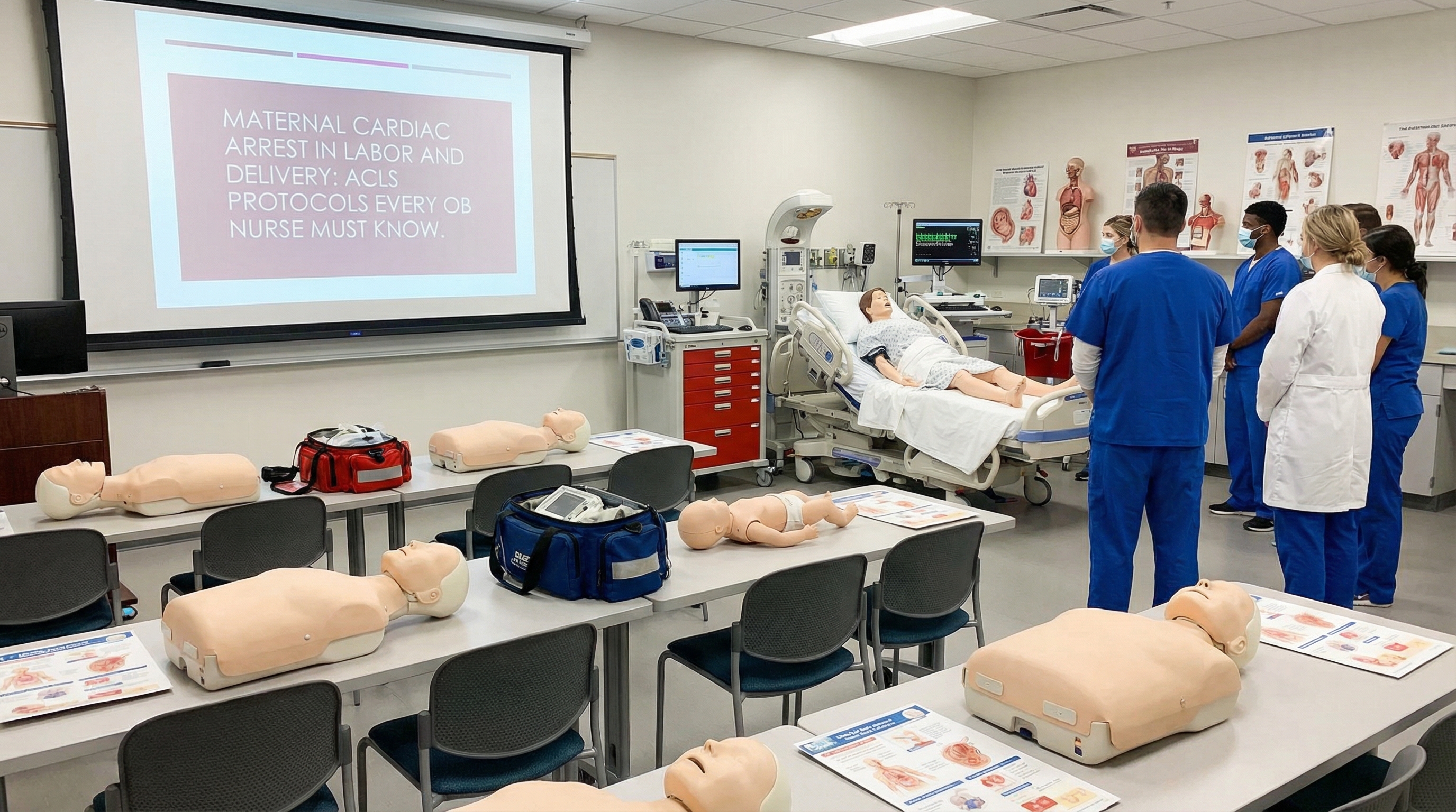
The underlying structure of post-arrest care follows the standard cardiac arrest algorithm, with obstetric modifications layered on top. Teams should debrief after every maternal code — both actual events and simulations — to identify what went well and what needs improvement. According to research published in JAMA Network Open on obstetric life support education, simulation-based training for maternal cardiac arrest significantly improves team performance, response times, and individual confidence in executing protocol steps — making regular drills a patient safety imperative, not an optional exercise.
OB nurses who participate in regular mock codes perform measurably better during actual maternal arrest events — not because the training adds new knowledge in the moment, but because it automates the right responses so that stress and adrenaline do not override them. This is not a theory; it is backed by evidence. A study cited by the Alliance for Innovation on Maternal Health (AIM) program on cardiac conditions in obstetric care demonstrates measurable improvement in team-based outcomes when obstetric units implement regular resuscitation simulation protocols.
Despite this evidence, many OB units run infrequent simulations, and many OB nurses hold ACLS certifications obtained from general training programs that do not include obstetric-specific content. Closing this gap is a patient safety imperative. Your ACLS certification is the foundation — but it requires you to actively apply the maternal modifications that standard courses may not cover in depth. Nurses who understand the modified resuscitation techniques for pregnant patients and practice them in their unit's simulation program are the ones who perform under pressure when it matters most.
The National Association of EMS Physicians resource on maternal cardiac arrest emphasizes that obstetric care teams should conduct detailed simulations incorporating the full range of interventions — LUD, PMCD decision timing, airway management, and team communication — not just individual skill stations. The entire team needs to practice the choreography of a maternal code together, not just know the steps individually.
For OB nurses who need to obtain or renew ACLS certification in a format that fits demanding shift schedules, online certification through board-certified emergency physicians offers a practical and affordable solution. Affordable ACLS provides ER physician-developed ACLS certification at just $99 — or $89 for recertification — with 100% self-paced online learning, immediate digital certification upon completion, and unlimited exam retakes. You can complete your certification between shifts without losing clinical hours, and then bring that knowledge directly to your unit's simulation and protocol training.
Keep this checklist accessible in your unit's code response documentation for rapid reference during an event:
Maternal cardiac arrest is one of the most demanding emergencies in all of healthcare. It demands technical precision, team coordination, time-critical decision-making, and the ability to execute complex protocols under the highest possible stress — all while managing two patients simultaneously. No other cardiac arrest scenario combines this level of physiologic complexity with this degree of emotional weight.
The OB nurses who perform best in these moments share one characteristic: they prepared before the emergency happened. They know their ACLS algorithms with confidence. They understand the obstetric modifications and have practiced them in simulation. They know when to call for PMCD and how to advocate clearly for its timely initiation. They have rehearsed their team roles and communication scripts enough times that the right actions come automatically when they are needed most.
Building that foundation starts with solid ACLS certification and a commitment to applying it in the obstetric context. Whether you are obtaining your certification for the first time or renewing credentials that are about to expire, keeping your knowledge current is the single most important step you can take for your patients. The L&D unit is not where you want to be improvising — it is where you want to be executing a plan you have already fully internalized.
Affordable ACLS offers 100% online, self-paced ACLS certification developed by board-certified ER physicians, starting at just $99 with immediate digital certification, unlimited retakes, and a money-back guarantee. Renew or certify on your schedule — and bring that expertise to every shift in labor and delivery.
.jpg)