ACLS Blogs

ACLS Rhythm Recognition Cheat Sheet: A Printable Quick Reference

A Quick-Reference Cheat Sheet, Not a Tutorial

If you already understand the "why" behind ACLS rhythms and just need to confirm what you are looking at on the monitor in ten seconds or less, this cheat sheet is built for you. Every entry below follows the same three-part format: identifying features, whether the rhythm is shockable or not, and the first-line response. No lengthy explanations, no backstory, just the facts you need to confirm a rhythm and move.


If you are still building your foundation in rhythm recognition, or you want the deeper explanation of what each waveform represents and why it behaves the way it does, start with our beginner's guide to identifying core ACLS heart rhythms first. That article teaches the concepts. This one is meant to sit next to your monitor, printed out or pulled up on your phone, while you are actively working a code or drilling for your next recertification exam.


How to Use This Cheat Sheet

Each rhythm below is broken into the same three fields so you can scan for exactly what you need under pressure: Identifying Features (what you see on the strip), Shockable Status (whether defibrillation is indicated), and First-Line Response (the immediate action per current AHA ACLS guidelines). Print this page, laminate it, or save it to your phone's home screen. It is meant as a fast-recall reference for providers who are already certified or currently studying, not a substitute for full ACLS training or your facility's protocols, which always take precedence in a real event.

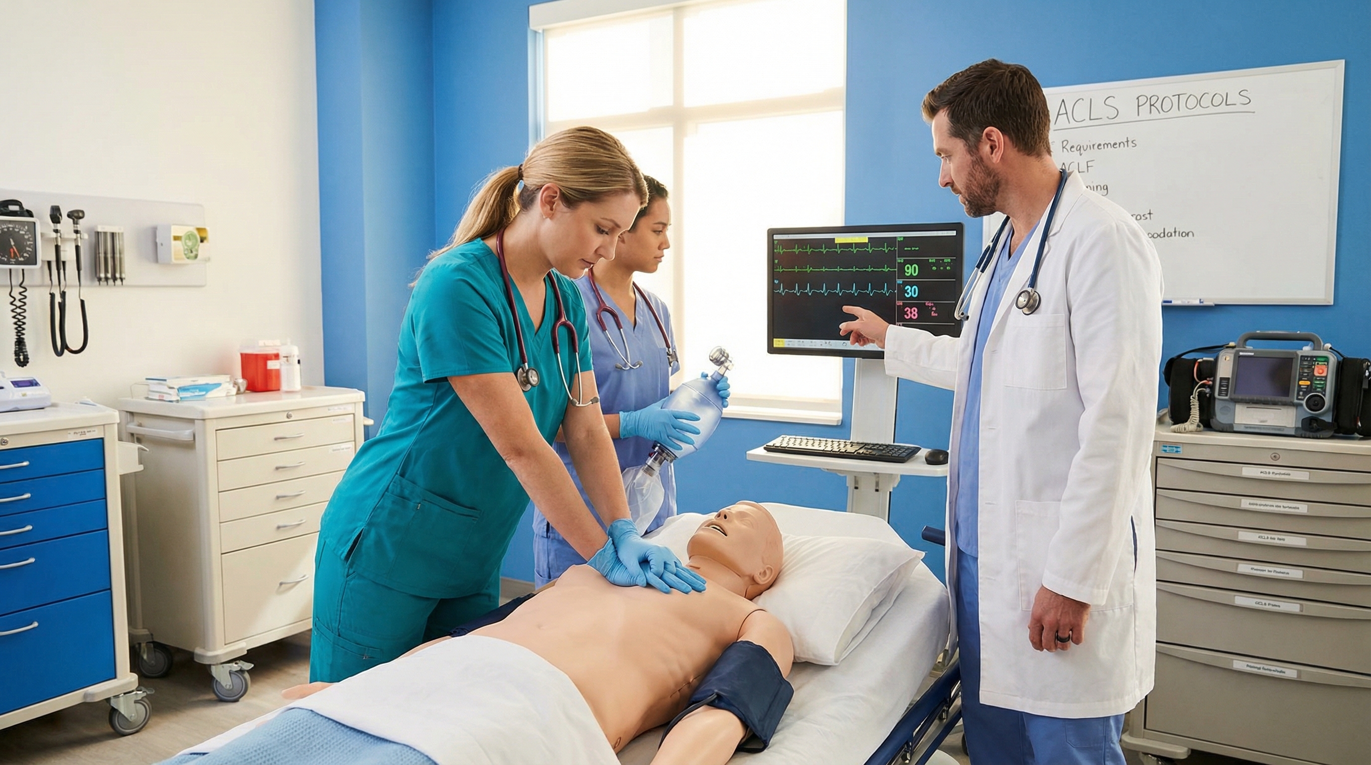
Healthcare providers reviewing a cardiac rhythm strip on a bedside monitor


Shockable vs. Non-Shockable: The Big Picture First

Before you drill into individual rhythms, anchor yourself to the single most important branch point in the cardiac arrest algorithm: is this rhythm shockable or not? Everything else, drug timing, compression cycles, and reassessment intervals, flows from that first decision. The current AHA Adult Cardiac Arrest Algorithm builds its entire structure around this same fork in the road, which is why it is worth memorizing before anything else on this page.


Shockable Rhythms (Defibrillate)

  • Ventricular Fibrillation (VF)
  • Pulseless Ventricular Tachycardia (pVT)

Non-Shockable Rhythms (No Defibrillation)

  • Asystole
  • Pulseless Electrical Activity (PEA)

For a full breakdown of why these two non-shockable rhythms get treated so differently despite both lacking a pulse, see our guide to the key differences between asystole and PEA.


The Core Cardiac Arrest Rhythms

These four rhythms make up the entire adult cardiac arrest algorithm. Every pulseless patient you encounter falls into one of these categories, and each one demands a different first move.


Ventricular Fibrillation (VF)

  • Chaotic, disorganized waveform with no identifiable P waves, QRS complexes, or T waves
  • Amplitude may be coarse (larger, more organized-looking) or fine (low amplitude, closer to asystole)
  • No coordinated ventricular contraction, so there is no pulse and no cardiac output
  • Rate and rhythm are not measurable because there is no true "beat" to count

Shockable Status: Yes. VF is defibrillated immediately upon recognition.


  • Deliver one shock as soon as the rhythm is confirmed, then resume chest compressions immediately without pausing to check a pulse
  • Continue high-quality CPR for two minutes before the next rhythm check
  • Establish IV or IO access and give epinephrine after the first defibrillation attempt fails to convert the rhythm
  • Consider amiodarone or lidocaine if VF persists after defibrillation, CPR, and vasopressor administration

For a deeper look at how VF and pVT compare on the strip and why both call for the same immediate response, see our guide to shockable rhythms: ventricular tachycardia and fibrillation.


Pulseless Ventricular Tachycardia (pVT)

  • Wide QRS complexes (greater than 0.12 seconds) occurring in a fast, regular pattern
  • Unlike VF, pVT has an organized, repeating waveform, but it still produces no palpable pulse
  • Rate is typically well above 100 beats per minute, often 150 to 250
  • Can appear monomorphic (uniform QRS shape) or polymorphic (varying QRS shape, which raises suspicion for torsades)

Shockable Status: Yes. Treated identically to VF in the pulseless patient.


  • Defibrillate immediately, then resume CPR for two minutes
  • Follow the same epinephrine and antiarrhythmic sequence used for VF
  • Reassess rhythm and pulse every two minutes, minimizing interruptions to compressions

Asystole

  • Flat line with no discernible electrical activity
  • Always confirm in more than one lead before calling it asystole, since a flat line can also indicate a disconnected lead or a fine VF that appears flat in a single view
  • No P waves, no QRS complexes, no measurable rate

Shockable Status: No. Defibrillation is never indicated for a true flat line.


  • Continue high-quality CPR without interruption for defibrillation
  • Give epinephrine as soon as feasible, then repeat every three to five minutes
  • Search aggressively for reversible causes, since asystole is often the end result of an untreated underlying problem rather than a primary rhythm disorder
  • Consider terminating resuscitation per your facility's protocol if asystole persists despite adequate CPR, medications, and correction of reversible causes

Pulseless Electrical Activity (PEA)

  • Organized electrical activity on the monitor, sometimes even a near-normal-looking QRS complex, but no palpable pulse
  • Rhythm can appear narrow or wide, fast or slow, which is what makes PEA tricky to recognize by strip alone
  • The defining feature is the mismatch between organized electrical activity and the complete absence of mechanical contraction strong enough to produce a pulse

Shockable Status: No. The problem is mechanical or metabolic, not electrical, so a shock will not help.

  • Continue CPR and give epinephrine as soon as feasible
  • Work through the Hs and Ts systematically, since PEA is almost always caused by an identifiable and often reversible underlying condition
  • Use point-of-care ultrasound or capnography where available to help narrow down the likely cause while compressions continue
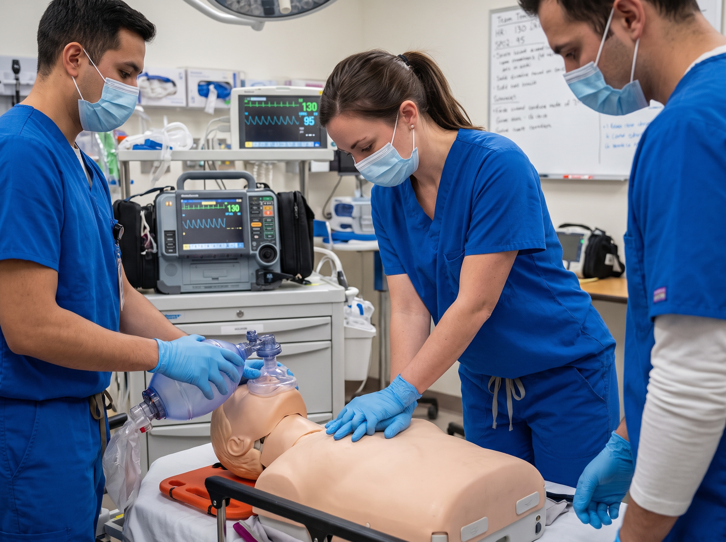
Code team performing CPR during a simulated cardiac arrest with monitor showing rhythm strip

Unstable and Symptomatic Rhythms (Pulse Present)

Not every rhythm on this sheet involves a pulseless patient. These next rhythms have a pulse, but the patient is often unstable and deteriorating quickly. Recognizing them fast prevents a slower problem from becoming a cardiac arrest.


Symptomatic Bradycardia

  • Heart rate typically under 50 beats per minute in a patient showing signs of poor perfusion
  • Watch for hypotension, altered mental status, chest pain, acute heart failure, or signs of shock accompanying the slow rate
  • Rhythm strip alone does not tell you whether to treat. A slow rate with a stable patient is monitored, not medicated

Shockable Status: Not applicable. Bradycardia with a pulse is never defibrillated.


  • Give atropine first if the patient is symptomatic
  • Move to transcutaneous pacing or a dopamine or epinephrine infusion if atropine is ineffective
  • Identify and treat the underlying cause, since bradycardia is frequently a downstream sign of hypoxia, medication effect, or an electrolyte problem

For a full walkthrough of dosing thresholds and when to escalate to pacing, see our guide to symptomatic bradycardia causes and treatment.


Unstable Tachycardia (SVT, Atrial Fibrillation with RVR, Monomorphic VT with a Pulse)

  • Heart rate generally above 150 beats per minute in a patient with a pulse
  • Narrow, regular QRS suggests SVT. Narrow, irregular QRS suggests atrial fibrillation or flutter with rapid ventricular response
  • Wide, regular QRS in a patient with a pulse should be treated as monomorphic VT until proven otherwise
  • Instability signs include hypotension, altered mental status, signs of shock, ischemic chest discomfort, or acute heart failure

Shockable Status: Not defibrillated. If unstable, this calls for synchronized cardioversion, not a defibrillation shock.


  • If unstable, prepare for immediate synchronized cardioversion
  • If stable, further evaluate the QRS width and regularity to guide medication choice, such as adenosine for regular narrow-complex SVT
  • Reassess frequently, since an unstable tachycardia can deteriorate into a pulseless rhythm quickly if untreated

Special Rhythm Considerations

These rhythms do not always fit neatly into the categories above, but providers are expected to recognize them quickly because the correct response differs meaningfully from a standard tachycardia or bradycardia.


AV Blocks: Which Ones Need a Pacer

  • First-degree AV block: Prolonged PR interval greater than 0.20 seconds, but every P wave still conducts to a QRS. Usually benign and monitored, not treated
  • Second-degree AV block, Type I (Wenckebach): Progressive PR prolongation until a QRS is dropped. Often stable and rarely requires pacing unless the patient is symptomatic
  • Second-degree AV block, Type II: Constant PR interval with intermittently dropped QRS complexes. Considered higher-risk and often progresses to complete block
  • Third-degree (complete) AV block: Complete dissociation between P waves and QRS complexes, each firing independently. Frequently requires transcutaneous or transvenous pacing

Shockable Status: No AV block is ever defibrillated. Type II second-degree and third-degree blocks in a symptomatic patient are managed the same way as symptomatic bradycardia: atropine first, then pacing if the patient does not respond.


Atrial Fibrillation and Atrial Flutter

  • Atrial fibrillation: Irregularly irregular narrow-complex rhythm with no discernible P waves, replaced by fibrillatory baseline activity
  • Atrial flutter: Regular sawtooth-shaped flutter waves, often at a predictable atrial rate with variable conduction ratios to the ventricles

Shockable Status: No, unless the patient is unstable, in which case synchronized cardioversion (not defibrillation) is indicated.


  • If stable, focus on rate control and evaluating anticoagulation needs based on duration and risk factors
  • If unstable, prepare for synchronized cardioversion
  • Always reassess whether rapid ventricular response is the primary problem or a symptom of something else, such as sepsis, hypovolemia, or thyroid storm

Torsades de Pointes

  • A distinct polymorphic VT where the QRS complexes appear to twist around the baseline in amplitude and axis
  • Almost always associated with a prolonged QT interval on a prior baseline strip
  • Can be self-terminating and recurrent, or can degenerate into VF

Shockable Status: Yes if the patient is pulseless. Defibrillate exactly as you would for VF or pVT.


  • If pulseless, defibrillate immediately and follow the standard cardiac arrest sequence
  • Give IV magnesium sulfate promptly, since it is the first-line treatment for torsades regardless of the patient's measured magnesium level
  • Identify and remove the offending cause where possible, such as a QT-prolonging medication or an electrolyte disturbance

Research on magnesium sulfate for torsades has shown consistent suppression of the arrhythmia even when serum magnesium levels are already within normal range, according to a case report and review published in PMC describing successful management of refractory torsades guided by ionized magnesium monitoring.


How Rhythm Recognition Drives Your Next Move

The entire point of rhythm recognition under pressure is speed to the correct branch of the algorithm. Confirming shockable versus non-shockable in the first few seconds determines whether your next action is charging the defibrillator or continuing compressions while you push epinephrine. According to the 2024 International Consensus on Cardiopulmonary Resuscitation and Emergency Cardiovascular Care Science published through the American Heart Association, rhythm check timing and minimizing interruptions in chest compressions remain central priorities across current resuscitation science. These treatment recommendations are developed through the ongoing systematic review process coordinated by the International Liaison Committee on Resuscitation, the global body that evaluates the evidence behind the algorithm steps summarized on this page.


This is also why the initial rhythm matters for prognosis, not just for the immediate next step. A study on the prognostic implications of rhythm conversion during out-of-hospital cardiac arrest found that patients whose initial non-shockable rhythm converted to a shockable one during resuscitation had meaningfully different outcomes compared to those who stayed in a non-shockable rhythm throughout the event, underscoring why continuous reassessment every two minutes is not just procedural, it changes clinical decision-making in real time.


If you want the complete algorithm flow that ties every rhythm on this page to its corresponding treatment pathway, bookmark our ACLS algorithms hub, which walks through each decision point step by step alongside the rhythm strips discussed here.


Keeping This Cheat Sheet Useful in Real Codes and on Exam Day

A cheat sheet only helps if you can recall it without looking, which means the printed version should be a backup, not your primary study method. Pair this reference with active recall: cover the identifying features column and quiz yourself on shockable status and first-line response from memory, then check your answer against the sheet.


Rhythm recognition rarely shows up in isolation on your certification exam. It is almost always tested alongside medication selection and timing, so pairing this rhythm reference with our ACLS medications cheat sheet gives you both halves of the megacode equation in one study session. If you want to see exactly how these rhythms and medications come together under exam conditions, our station-by-station megacode walkthrough shows how a rhythm change mid-scenario should change your called-out actions in real time.


Building Rhythm Recall Into Your Certification

Rhythm recognition is not a skill you build once and keep forever. It fades between recertifications the same way any pattern-recognition skill does when it goes unused for months at a time. The most reliable way to keep it sharp is to revisit strips regularly, not just in the weeks before your card expires.


If your certification is due for renewal or you are preparing for your first ACLS card, our ACLS certification and recertification course was built by practicing emergency medicine physicians and covers every rhythm on this cheat sheet in full clinical context, with unlimited retakes if a topic does not click on the first pass. The course is self-paced, so you can move quickly through material you already know and slow down on the rhythms that still trip you up.


Frequently Asked Rhythm Recognition Questions

A few questions come up constantly during ACLS review sessions and megacode prep. Here are quick answers to the ones providers ask most.


What is the practical difference between VF and pVT if they are treated the same way?

Clinically, the treatment pathway is identical, defibrillate, resume CPR, reassess in two minutes. The distinction matters mainly for documentation and pattern recognition. VF has no organized waveform at all, while pVT shows a repeating, wide-complex pattern. Being able to tell them apart quickly helps you communicate clearly with your code team and supports accurate charting after the event.


How do you avoid mistaking fine VF for asystole?

Always confirm a flat-looking rhythm in at least two leads before calling asystole, and check that the gain on the monitor is turned up enough to catch low-amplitude fibrillatory waves. A lead that has come loose or a signal that is too small to see can both mimic a flat line. When in doubt, treat it as a non-shockable rhythm and keep compressions going while you troubleshoot the monitor.


Can PEA have a fast rate, a slow rate, or both?

Both. PEA is defined by the mismatch between organized electrical activity and the absence of a palpable pulse, not by any specific rate or QRS width. You can see PEA with a narrow, fast complex or a wide, slow one. That variability is exactly why the Hs and Ts workup matters so much here, the strip alone will not tell you what is causing the arrest.


When do you use synchronized cardioversion instead of defibrillation?

Synchronized cardioversion is used for an unstable patient who still has a pulse, such as unstable atrial fibrillation with rapid ventricular response or unstable monomorphic VT with a pulse. Defibrillation, an unsynchronized shock, is reserved for pulseless shockable rhythms like VF and pVT, or for polymorphic VT where synchronization is not reliable. Mixing these up is one of the more common megacode mistakes, so it is worth drilling the distinction until it is automatic.


Final Thoughts

Rhythm recognition is a perishable skill that rewards repetition far more than it rewards a single study session. Keep this cheat sheet within reach during shifts and study blocks, revisit the rhythms that give you the most trouble, and lean on the deeper guides linked throughout this page whenever a rhythm needs more explanation than a bullet list can offer.


ACLS Blogs

ACLS Rhythm Recognition Cheat Sheet: A Printable Quick Reference

A Quick-Reference Cheat Sheet, Not a Tutorial

If you already understand the "why" behind ACLS rhythms and just need to confirm what you are looking at on the monitor in ten seconds or less, this cheat sheet is built for you. Every entry below follows the same three-part format: identifying features, whether the rhythm is shockable or not, and the first-line response. No lengthy explanations, no backstory, just the facts you need to confirm a rhythm and move.


If you are still building your foundation in rhythm recognition, or you want the deeper explanation of what each waveform represents and why it behaves the way it does, start with our beginner's guide to identifying core ACLS heart rhythms first. That article teaches the concepts. This one is meant to sit next to your monitor, printed out or pulled up on your phone, while you are actively working a code or drilling for your next recertification exam.


How to Use This Cheat Sheet

Each rhythm below is broken into the same three fields so you can scan for exactly what you need under pressure: Identifying Features (what you see on the strip), Shockable Status (whether defibrillation is indicated), and First-Line Response (the immediate action per current AHA ACLS guidelines). Print this page, laminate it, or save it to your phone's home screen. It is meant as a fast-recall reference for providers who are already certified or currently studying, not a substitute for full ACLS training or your facility's protocols, which always take precedence in a real event.

Healthcare providers reviewing a cardiac rhythm strip on a bedside monitor


Shockable vs. Non-Shockable: The Big Picture First

Before you drill into individual rhythms, anchor yourself to the single most important branch point in the cardiac arrest algorithm: is this rhythm shockable or not? Everything else, drug timing, compression cycles, and reassessment intervals, flows from that first decision. The current AHA Adult Cardiac Arrest Algorithm builds its entire structure around this same fork in the road, which is why it is worth memorizing before anything else on this page.


Shockable Rhythms (Defibrillate)

  • Ventricular Fibrillation (VF)
  • Pulseless Ventricular Tachycardia (pVT)

Non-Shockable Rhythms (No Defibrillation)

  • Asystole
  • Pulseless Electrical Activity (PEA)

For a full breakdown of why these two non-shockable rhythms get treated so differently despite both lacking a pulse, see our guide to the key differences between asystole and PEA.


The Core Cardiac Arrest Rhythms

These four rhythms make up the entire adult cardiac arrest algorithm. Every pulseless patient you encounter falls into one of these categories, and each one demands a different first move.


Ventricular Fibrillation (VF)

  • Chaotic, disorganized waveform with no identifiable P waves, QRS complexes, or T waves
  • Amplitude may be coarse (larger, more organized-looking) or fine (low amplitude, closer to asystole)
  • No coordinated ventricular contraction, so there is no pulse and no cardiac output
  • Rate and rhythm are not measurable because there is no true "beat" to count

Shockable Status: Yes. VF is defibrillated immediately upon recognition.


  • Deliver one shock as soon as the rhythm is confirmed, then resume chest compressions immediately without pausing to check a pulse
  • Continue high-quality CPR for two minutes before the next rhythm check
  • Establish IV or IO access and give epinephrine after the first defibrillation attempt fails to convert the rhythm
  • Consider amiodarone or lidocaine if VF persists after defibrillation, CPR, and vasopressor administration

For a deeper look at how VF and pVT compare on the strip and why both call for the same immediate response, see our guide to shockable rhythms: ventricular tachycardia and fibrillation.


Pulseless Ventricular Tachycardia (pVT)

  • Wide QRS complexes (greater than 0.12 seconds) occurring in a fast, regular pattern
  • Unlike VF, pVT has an organized, repeating waveform, but it still produces no palpable pulse
  • Rate is typically well above 100 beats per minute, often 150 to 250
  • Can appear monomorphic (uniform QRS shape) or polymorphic (varying QRS shape, which raises suspicion for torsades)

Shockable Status: Yes. Treated identically to VF in the pulseless patient.


  • Defibrillate immediately, then resume CPR for two minutes
  • Follow the same epinephrine and antiarrhythmic sequence used for VF
  • Reassess rhythm and pulse every two minutes, minimizing interruptions to compressions

Asystole

  • Flat line with no discernible electrical activity
  • Always confirm in more than one lead before calling it asystole, since a flat line can also indicate a disconnected lead or a fine VF that appears flat in a single view
  • No P waves, no QRS complexes, no measurable rate

Shockable Status: No. Defibrillation is never indicated for a true flat line.


  • Continue high-quality CPR without interruption for defibrillation
  • Give epinephrine as soon as feasible, then repeat every three to five minutes
  • Search aggressively for reversible causes, since asystole is often the end result of an untreated underlying problem rather than a primary rhythm disorder
  • Consider terminating resuscitation per your facility's protocol if asystole persists despite adequate CPR, medications, and correction of reversible causes

Pulseless Electrical Activity (PEA)

  • Organized electrical activity on the monitor, sometimes even a near-normal-looking QRS complex, but no palpable pulse
  • Rhythm can appear narrow or wide, fast or slow, which is what makes PEA tricky to recognize by strip alone
  • The defining feature is the mismatch between organized electrical activity and the complete absence of mechanical contraction strong enough to produce a pulse

Shockable Status: No. The problem is mechanical or metabolic, not electrical, so a shock will not help.


  • Continue CPR and give epinephrine as soon as feasible
  • Work through the Hs and Ts systematically, since PEA is almost always caused by an identifiable and often reversible underlying condition
  • Use point-of-care ultrasound or capnography where available to help narrow down the likely cause while compressions continue
Code team performing CPR during a simulated cardiac arrest with monitor showing rhythm strip

Unstable and Symptomatic Rhythms (Pulse Present)

Not every rhythm on this sheet involves a pulseless patient. These next rhythms have a pulse, but the patient is often unstable and deteriorating quickly. Recognizing them fast prevents a slower problem from becoming a cardiac arrest.


Symptomatic Bradycardia

  • Heart rate typically under 50 beats per minute in a patient showing signs of poor perfusion
  • Watch for hypotension, altered mental status, chest pain, acute heart failure, or signs of shock accompanying the slow rate
  • Rhythm strip alone does not tell you whether to treat. A slow rate with a stable patient is monitored, not medicated

Shockable Status: Not applicable. Bradycardia with a pulse is never defibrillated.


  • Give atropine first if the patient is symptomatic
  • Move to transcutaneous pacing or a dopamine or epinephrine infusion if atropine is ineffective
  • Identify and treat the underlying cause, since bradycardia is frequently a downstream sign of hypoxia, medication effect, or an electrolyte problem

For a full walkthrough of dosing thresholds and when to escalate to pacing, see our guide to symptomatic bradycardia causes and treatment.


Unstable Tachycardia (SVT, Atrial Fibrillation with RVR, Monomorphic VT with a Pulse)

  • Heart rate generally above 150 beats per minute in a patient with a pulse
  • Narrow, regular QRS suggests SVT. Narrow, irregular QRS suggests atrial fibrillation or flutter with rapid ventricular response
  • Wide, regular QRS in a patient with a pulse should be treated as monomorphic VT until proven otherwise
  • Instability signs include hypotension, altered mental status, signs of shock, ischemic chest discomfort, or acute heart failure

Shockable Status: Not defibrillated. If unstable, this calls for synchronized cardioversion, not a defibrillation shock.


  • If unstable, prepare for immediate synchronized cardioversion
  • If stable, further evaluate the QRS width and regularity to guide medication choice, such as adenosine for regular narrow-complex SVT
  • Reassess frequently, since an unstable tachycardia can deteriorate into a pulseless rhythm quickly if untreated

Special Rhythm Considerations

These rhythms do not always fit neatly into the categories above, but providers are expected to recognize them quickly because the correct response differs meaningfully from a standard tachycardia or bradycardia.


AV Blocks: Which Ones Need a Pacer

  • First-degree AV block: Prolonged PR interval greater than 0.20 seconds, but every P wave still conducts to a QRS. Usually benign and monitored, not treated
  • Second-degree AV block, Type I (Wenckebach): Progressive PR prolongation until a QRS is dropped. Often stable and rarely requires pacing unless the patient is symptomatic
  • Second-degree AV block, Type II: Constant PR interval with intermittently dropped QRS complexes. Considered higher-risk and often progresses to complete block
  • Third-degree (complete) AV block: Complete dissociation between P waves and QRS complexes, each firing independently. Frequently requires transcutaneous or transvenous pacing

Shockable Status: No AV block is ever defibrillated. Type II second-degree and third-degree blocks in a symptomatic patient are managed the same way as symptomatic bradycardia: atropine first, then pacing if the patient does not respond.


Atrial Fibrillation and Atrial Flutter

  • Atrial fibrillation: Irregularly irregular narrow-complex rhythm with no discernible P waves, replaced by fibrillatory baseline activity
  • Atrial flutter: Regular sawtooth-shaped flutter waves, often at a predictable atrial rate with variable conduction ratios to the ventricles

Shockable Status: No, unless the patient is unstable, in which case synchronized cardioversion (not defibrillation) is indicated.


  • If stable, focus on rate control and evaluating anticoagulation needs based on duration and risk factors
  • If unstable, prepare for synchronized cardioversion
  • Always reassess whether rapid ventricular response is the primary problem or a symptom of something else, such as sepsis, hypovolemia, or thyroid storm

Torsades de Pointes

  • A distinct polymorphic VT where the QRS complexes appear to twist around the baseline in amplitude and axis
  • Almost always associated with a prolonged QT interval on a prior baseline strip
  • Can be self-terminating and recurrent, or can degenerate into VF

Shockable Status: Yes if the patient is pulseless. Defibrillate exactly as you would for VF or pVT.


  • If pulseless, defibrillate immediately and follow the standard cardiac arrest sequence
  • Give IV magnesium sulfate promptly, since it is the first-line treatment for torsades regardless of the patient's measured magnesium level
  • Identify and remove the offending cause where possible, such as a QT-prolonging medication or an electrolyte disturbance

Research on magnesium sulfate for torsades has shown consistent suppression of the arrhythmia even when serum magnesium levels are already within normal range, according to a case report and review published in PMC describing successful management of refractory torsades guided by ionized magnesium monitoring.


How Rhythm Recognition Drives Your Next Move

The entire point of rhythm recognition under pressure is speed to the correct branch of the algorithm. Confirming shockable versus non-shockable in the first few seconds determines whether your next action is charging the defibrillator or continuing compressions while you push epinephrine. According to the 2024 International Consensus on Cardiopulmonary Resuscitation and Emergency Cardiovascular Care Science published through the American Heart Association, rhythm check timing and minimizing interruptions in chest compressions remain central priorities across current resuscitation science. These treatment recommendations are developed through the ongoing systematic review process coordinated by the International Liaison Committee on Resuscitation, the global body that evaluates the evidence behind the algorithm steps summarized on this page.


This is also why the initial rhythm matters for prognosis, not just for the immediate next step. A study on the prognostic implications of rhythm conversion during out-of-hospital cardiac arrest found that patients whose initial non-shockable rhythm converted to a shockable one during resuscitation had meaningfully different outcomes compared to those who stayed in a non-shockable rhythm throughout the event, underscoring why continuous reassessment every two minutes is not just procedural, it changes clinical decision-making in real time.


If you want the complete algorithm flow that ties every rhythm on this page to its corresponding treatment pathway, bookmark our ACLS algorithms hub, which walks through each decision point step by step alongside the rhythm strips discussed here.


Keeping This Cheat Sheet Useful in Real Codes and on Exam Day

A cheat sheet only helps if you can recall it without looking, which means the printed version should be a backup, not your primary study method. Pair this reference with active recall: cover the identifying features column and quiz yourself on shockable status and first-line response from memory, then check your answer against the sheet.


Rhythm recognition rarely shows up in isolation on your certification exam. It is almost always tested alongside medication selection and timing, so pairing this rhythm reference with our ACLS medications cheat sheet gives you both halves of the megacode equation in one study session. If you want to see exactly how these rhythms and medications come together under exam conditions, our station-by-station megacode walkthrough shows how a rhythm change mid-scenario should change your called-out actions in real time.


Building Rhythm Recall Into Your Certification

Rhythm recognition is not a skill you build once and keep forever. It fades between recertifications the same way any pattern-recognition skill does when it goes unused for months at a time. The most reliable way to keep it sharp is to revisit strips regularly, not just in the weeks before your card expires.


If your certification is due for renewal or you are preparing for your first ACLS card, our ACLS certification and recertification course was built by practicing emergency medicine physicians and covers every rhythm on this cheat sheet in full clinical context, with unlimited retakes if a topic does not click on the first pass. The course is self-paced, so you can move quickly through material you already know and slow down on the rhythms that still trip you up.


Frequently Asked Rhythm Recognition Questions

A few questions come up constantly during ACLS review sessions and megacode prep. Here are quick answers to the ones providers ask most.


What is the practical difference between VF and pVT if they are treated the same way?

Clinically, the treatment pathway is identical, defibrillate, resume CPR, reassess in two minutes. The distinction matters mainly for documentation and pattern recognition. VF has no organized waveform at all, while pVT shows a repeating, wide-complex pattern. Being able to tell them apart quickly helps you communicate clearly with your code team and supports accurate charting after the event.


How do you avoid mistaking fine VF for asystole?

Always confirm a flat-looking rhythm in at least two leads before calling asystole, and check that the gain on the monitor is turned up enough to catch low-amplitude fibrillatory waves. A lead that has come loose or a signal that is too small to see can both mimic a flat line. When in doubt, treat it as a non-shockable rhythm and keep compressions going while you troubleshoot the monitor.


Can PEA have a fast rate, a slow rate, or both?

Both. PEA is defined by the mismatch between organized electrical activity and the absence of a palpable pulse, not by any specific rate or QRS width. You can see PEA with a narrow, fast complex or a wide, slow one. That variability is exactly why the Hs and Ts workup matters so much here, the strip alone will not tell you what is causing the arrest.


When do you use synchronized cardioversion instead of defibrillation?

Synchronized cardioversion is used for an unstable patient who still has a pulse, such as unstable atrial fibrillation with rapid ventricular response or unstable monomorphic VT with a pulse. Defibrillation, an unsynchronized shock, is reserved for pulseless shockable rhythms like VF and pVT, or for polymorphic VT where synchronization is not reliable. Mixing these up is one of the more common megacode mistakes, so it is worth drilling the distinction until it is automatic.


Final Thoughts

Rhythm recognition is a perishable skill that rewards repetition far more than it rewards a single study session. Keep this cheat sheet within reach during shifts and study blocks, revisit the rhythms that give you the most trouble, and lean on the deeper guides linked throughout this page whenever a rhythm needs more explanation than a bullet list can offer.


blogs

More articles for you

all articles

Start Today!

Get certified at your own pace, on your schedule, from the comfort of your home.