Every code starts the same way: a patient loses their pulse, an alarm sounds, and every provider in the room turns to the monitor. What happens in the next ten seconds — correctly identifying what that waveform is telling you — determines everything that follows. Rhythm recognition is not just a test question. It is the single most time-critical clinical skill you exercise during a cardiac arrest.
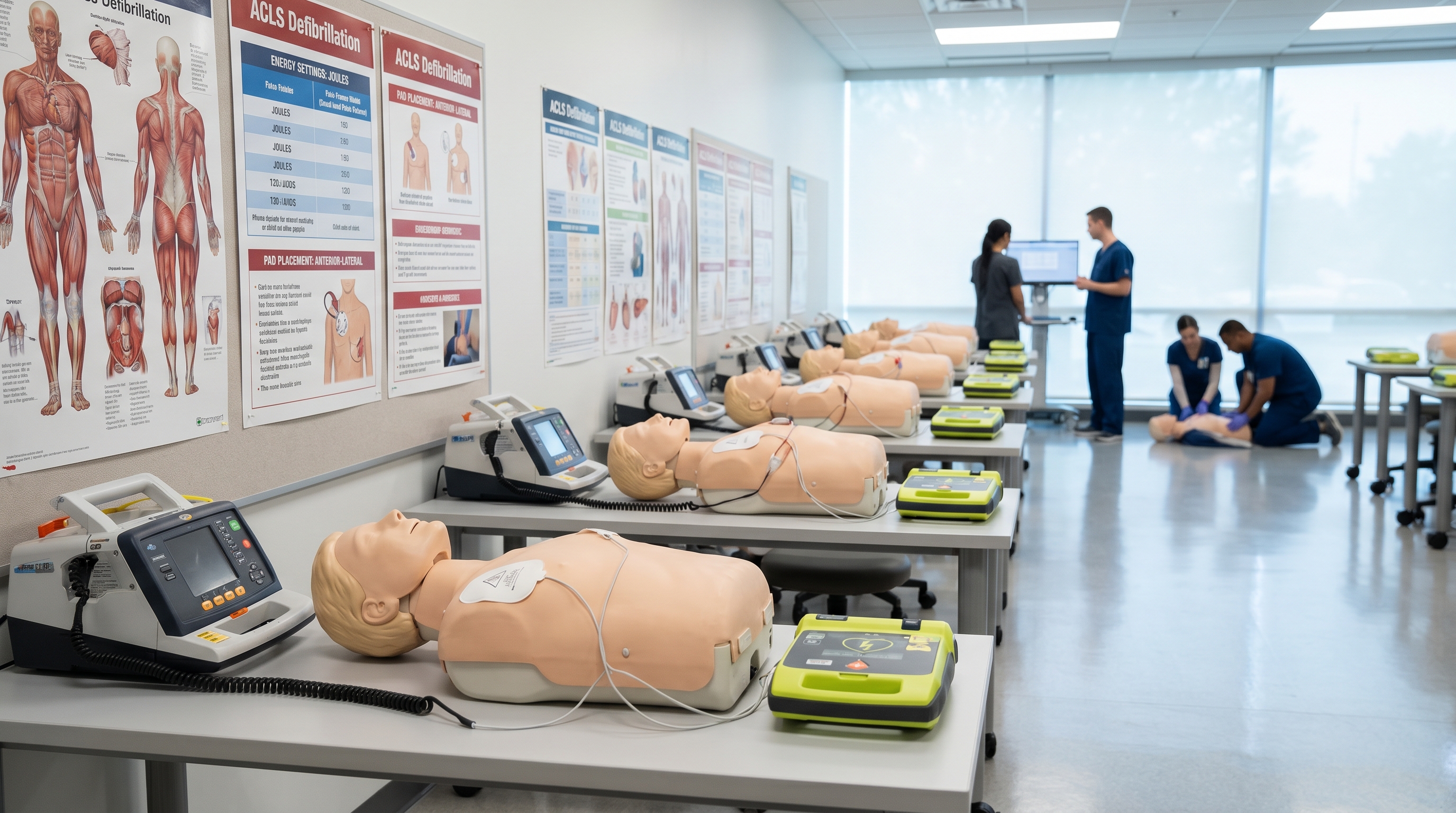
If you are relatively new to Advanced Cardiovascular Life Support or are preparing for your first ACLS certification, the monitor can feel overwhelming. Waveforms scroll across the screen, numbers flash, and the team is waiting on a decision. This guide is written peer-to-peer, from emergency physicians who have stood at that bedside, to help you build a reliable mental framework for identifying the core ACLS rhythms before the pressure is on.

You do not need to memorize every rare dysrhythmia to function effectively in a code. You need to master the rhythms that drive ACLS algorithms — the ones that determine whether you shock, push epinephrine, pace, or keep doing CPR. Understanding these rhythms deeply will make every other aspect of ACLS click into place.
Before diving into specific rhythms, it helps to speak the same language as the monitor. A standard ECG tracing shows electrical activity moving through the heart in a predictable sequence. The key components you need to recognize are:
When you approach any rhythm strip, run through five questions systematically: What is the rate? Is the rhythm regular or irregular? Are there P waves? Is the PR interval normal? Is the QRS narrow or wide? These five questions will route you to the correct rhythm category every single time. According to current ACLS ECG interpretation guidelines, this systematic approach is the clinical standard taught to providers at every level.
The most critical distinction in ACLS rhythm interpretation is shockable versus non-shockable. Shockable rhythms — ventricular fibrillation and pulseless ventricular tachycardia — are chaotic electrical states that can be terminated with a properly timed electrical shock. Non-shockable rhythms, by contrast, involve either no meaningful electrical activity or organized electrical activity without effective mechanical output. Shocking a non-shockable rhythm accomplishes nothing and wastes precious time.
Ventricular fibrillation is the cardiac arrest rhythm that defibrillation was designed to treat. When you see VF on the monitor, the ventricles are not contracting — they are quivering chaotically, driven by hundreds of disorganized electrical impulses firing simultaneously. There is no coordinated pump action, no cardiac output, and no pulse.
On the monitor, VF appears as a completely chaotic, disorganized waveform with no identifiable P waves, QRS complexes, or T waves. There is no regular pattern whatsoever. The amplitude can range from coarse (large, irregular waves) to fine (small, subtle waves that can be mistaken for artifact or even asystole). Coarse VF is generally more responsive to defibrillation and indicates a more recent onset.
The treatment pathway is immediate: high-quality CPR while the defibrillator charges, one shock, then two minutes of CPR before re-checking the rhythm. Epinephrine is administered every 3–5 minutes, and amiodarone or lidocaine is considered for shock-refractory VF. For a deeper clinical review of ventricular fibrillation mechanisms and treatment, see this comprehensive guide on understanding ventricular fibrillation causes, symptoms, and treatment.
Ventricular tachycardia (VT) can exist on a spectrum. When it is monomorphic and the patient has a pulse with stable blood pressure, it is a tachyarrhythmia requiring a different management approach. But pulseless VT — ventricular tachycardia with no palpable pulse — is a cardiac arrest rhythm treated identically to VF.
On the monitor, pulseless VT appears as a wide, regular, rapid rhythm — typically greater than 100 beats per minute and often 150–250 bpm. The QRS complexes are wide (greater than 0.12 seconds) and bizarre-looking compared to a normal sinus rhythm. Critically, there are no identifiable P waves preceding the QRS complexes.
The key clinical point: the monitor shows VT but the patient has no pulse. This is what separates pVT from stable VT. Always check for a pulse before treating. If no pulse is present, follow the shockable cardiac arrest algorithm. Providers who need a deeper reference on wide complex tachycardias should review this detailed clinical guide on understanding wide complex tachycardias.
According to StatPearls published by the NIH, PEA and asystole together account for approximately 81% of initial in-hospital cardiac arrest rhythms in the United States. This means the majority of codes you will run are non-shockable, making these rhythms equally critical to master. Survival rates for non-shockable rhythms are significantly lower than for shockable rhythms, which underscores the importance of high-quality CPR and rapid identification of reversible causes.

Asystole is the absence of all cardiac electrical activity. Clinically, it is what most people picture when they think of cardiac arrest — the flatline. On the monitor, asystole appears as a nearly flat, isoelectric line with no discernible waveform activity.
Before calling a rhythm asystole, always confirm in two leads. Artifact or a loose lead can mimic asystole, and the last thing you want to do is treat a patient in VF as if they are in asystole. If VF is even a possibility, treat it as VF and defibrillate.
Asystole is not shockable. The treatment is continuous high-quality CPR with epinephrine every 3–5 minutes, combined with an aggressive search for reversible causes using the H's and T's framework. Survival rates for asystole are very poor — published data suggests approximately 1.5% survival to 30 days when asystole is the initial rhythm — which is why early recognition of the H's and T's and rapid reversal of any correctable cause is paramount.
Pulseless electrical activity is in many ways the most conceptually challenging of the four core ACLS rhythms. Unlike VF, VT, and asystole, PEA is defined not by what the rhythm looks like, but by the mismatch between the electrical activity on the monitor and the absence of mechanical output. In PEA, there is organized (or semi-organized) electrical activity on the monitor, but the patient has no pulse and no effective cardiac output.
The rhythm on the monitor during PEA can look like almost anything — sinus rhythm, sinus tachycardia, a bradycardia, even something that looks like a relatively normal ECG. The defining clinical feature is the absence of a palpable pulse. This is why pulse checks are non-negotiable at every rhythm assessment in a code.
PEA is almost always caused by a reversible condition. The H's and T's framework was designed largely with PEA in mind: Hypovolemia, Hypoxia, Hydrogen ion (acidosis), Hypo/Hyperkalemia, Hypothermia, Tension pneumothorax, Tamponade, Toxins, Thrombosis (pulmonary), and Thrombosis (coronary). Treatment is high-quality CPR, epinephrine every 3–5 minutes, and rapid identification and correction of the underlying cause. For a thorough clinical reference, review this guide on understanding pulseless electrical activity causes and treatment.
Not every dangerous rhythm involves cardiac arrest. Symptomatic bradycardia — a heart rate below 60 bpm accompanied by hemodynamic instability, altered mental status, chest pain, or signs of shock — requires rapid recognition and treatment on its own algorithm track.
On the monitor, sinus bradycardia looks like a normal sinus rhythm but with a rate below 60. P waves are present, QRS complexes follow each P wave normally, and the overall morphology is unremarkable — except for the dangerously slow rate. The critical question is always: is the patient symptomatic? A heart rate of 48 in a trained endurance athlete sleeping comfortably is entirely benign. The same heart rate in a patient who is diaphoretic, hypotensive, and barely conscious is an emergency.
The ACLS bradycardia algorithm begins with atropine 0.5 mg IV, repeatable up to a total of 3 mg. If atropine fails to restore adequate rate and the patient remains symptomatic, transcutaneous pacing is the next step. For symptomatic bradycardia that is unresponsive to medications and pacing, transvenous pacing may ultimately be required. For a deeper dive into bradycardia presentation and treatment, this clinical article on understanding symptomatic bradycardia is an excellent reference.
Tachyarrhythmias on the ACLS monitor fall into two broad categories: stable (the patient is compensating) and unstable (the rapid rate is causing hemodynamic compromise). The first step in any tachyarrhythmia is always: is the patient stable or unstable? Unstable tachycardia — regardless of the specific rhythm — gets synchronized cardioversion immediately. Stable tachycardia gets a more deliberate diagnostic and treatment approach.
Atrial fibrillation is the most common sustained cardiac arrhythmia you will encounter in clinical practice, and it is highly prevalent in acutely ill and hospitalized patients. On the monitor, AFib has two defining features: the absence of distinct P waves (replaced by a chaotic, irregular baseline often described as fibrillatory waves) and an irregularly irregular ventricular rate. There is no consistent P-to-P or R-to-R interval.
The ventricular rate in AFib can range widely — from a controlled rate of 60–100 to a rapid ventricular response above 150. If the rate is rapid and the patient shows signs of hemodynamic compromise (hypotension, altered mental status, acute chest pain, signs of pulmonary edema), synchronized cardioversion is indicated. If the patient is stable, rate control and anticoagulation considerations come into play. For detailed reading on atrial fibrillation pathophysiology and management, see this guide on understanding atrial fibrillation causes, symptoms, and treatments.
Supraventricular tachycardia is a broad category encompassing any tachycardia with a circuit originating above the bundle of His. In the practical ACLS context, SVT most often refers to AVNRT (AV nodal reentrant tachycardia) — a regular, narrow complex tachycardia with rates typically between 150–250 bpm. The QRS complexes are narrow and regular, and P waves, if visible at all, may be buried in or just after the QRS complex.
In a stable patient, the initial treatment for SVT is vagal maneuvers followed by adenosine if vagal maneuvers fail. Adenosine 6 mg IV rapid push, followed by 12 mg if needed, works by transiently blocking AV node conduction and breaking the reentrant circuit. In an unstable patient, synchronized cardioversion is the immediate intervention. According to clinical guidelines on rhythm-based management, rapid identification of rhythm stability is the pivotal decision point that shapes the entire treatment path.
Atrioventricular blocks represent progressive failure of conduction through the AV node. On the ACLS monitor, you need to recognize three degrees of AV block and understand which ones require urgent intervention.
First-degree AV block is a prolonged PR interval (greater than 0.20 seconds) with every P wave followed by a QRS. It is generally benign and does not require ACLS-level treatment on its own.
Second-degree AV block Type I (Wenckebach) shows a progressively lengthening PR interval until a P wave is blocked entirely and no QRS follows. The cycle then resets. This rhythm is often well-tolerated but should be monitored closely. Second-degree Type II is more dangerous: the PR interval is constant, but beats are periodically dropped without warning. This pattern can degenerate into complete heart block and often requires pacing.
Third-degree AV block (complete heart block) is a true emergency. The atria and ventricles are firing completely independently. P waves march out at their own rate, QRS complexes march out at their own escape rate, and there is no relationship between them. The PR interval appears random because there is no conduction between the two. If symptomatic, this requires immediate transcutaneous pacing. For a comprehensive review of the AV block spectrum, this guide on understanding atrioventricular blocks covers all three degrees in clinical depth.
When adrenaline is running high and a team is watching you, having a systematic framework prevents tunnel vision and missed diagnoses. Emergency physicians and resuscitation experts consistently recommend the same five-step approach to rhythm analysis:
Providers who apply a systematic approach to rhythm analysis make faster and more accurate treatment decisions — a critical advantage when every second of cardiac arrest reduces the probability of neurologically intact survival. Research published through PMC on ECG interpretation proficiency confirms that structured training and systematic approaches significantly improve accuracy among healthcare providers, with the greatest gains seen in providers who practice regularly under supervised conditions.
Beyond the five-step approach, always contextualize the rhythm with the patient's clinical presentation. A wide complex tachycardia in a 65-year-old with a known history of heart disease and an ischemic cardiomyopathy is VT until proven otherwise. The same rhythm in a 25-year-old with Wolff-Parkinson-White may be SVT with aberrant conduction. The monitor tells you the electrical story; the patient tells you the clinical one.
For PEA and asystole especially — but relevant to any refractory cardiac arrest — the H's and T's provide a systematic checklist of reversible causes. Committing these to memory is a core ACLS competency that can directly change a patient's outcome:
Rhythm recognition is a skill that develops through repetition, not memorization. Reading about waveforms is the starting point, but the path to true fluency comes from looking at hundreds of rhythm strips and committing the visual patterns to long-term memory. Here are the strategies that emergency physicians and experienced ACLS educators consistently recommend:
The latest 2025 AHA ACLS guideline updates continue to reinforce rhythm-guided resuscitation as the centerpiece of effective cardiac arrest management. Staying current with guideline updates is part of your ongoing professional responsibility as an ACLS provider — certification is not a one-time event but a commitment to maintaining clinical readiness.
Understanding heart rhythms intellectually is step one. Putting that knowledge into practice within a structured, guideline-compliant curriculum is what earns you an ACLS certification that your employer will accept and that will serve you when it counts. For busy healthcare providers — nurses, physicians, paramedics, respiratory therapists — the traditional in-person ACLS course is not always practical. Schedules are unpredictable, training centers have limited seat availability, and spending an entire day away from clinical work or family is a real barrier.
That is exactly why Affordable ACLS was built by practicing Board Certified Emergency Medicine physicians who understand the demands placed on working clinicians. The platform delivers a fully online, self-paced ACLS certification built on current AHA and ILCOR guidelines — the same standards your hospital credentialing office requires. ACLS Certification is available at $99 and Recertification at $89, with a money-back guarantee if your employer does not accept the credential.
The course is completed at your own pace, on any device, with no time limits. You can work through the rhythm recognition modules during a slow overnight shift, finish the pharmacology sections on a lunch break, and complete your assessment when you are ready. Unlimited retakes at no additional charge mean you are not penalized for revisiting difficult material until you have truly mastered it. Your digital certificate is available for immediate download the moment you pass.
For nurses looking to leverage ACLS certification for career advancement, this guide on why nurses should choose online ACLS certification courses breaks down the professional and practical benefits. And if you are preparing for your first real code experience beyond just the certification exam, this guide on mental preparation for your first real code is essential reading for any new provider stepping into that role for the first time.
Reading a cardiac monitor accurately is one of those clinical skills that separates providers who are technically present during a code from those who are truly leading it. The rhythms covered in this guide — VF, pVT, asystole, PEA, symptomatic bradycardia, AFib, SVT, and AV blocks — form the complete core of what ACLS rhythm recognition requires. Each one has a distinct visual signature, a distinct clinical implication, and a distinct treatment pathway.
Master the five-step systematic approach. Understand the shockable versus non-shockable distinction at a reflex level. Know the H's and T's cold. Practice reading rhythm strips every chance you get. And back it all with a rigorous ACLS certification built on current guidelines. The investment you make now in rhythm mastery pays dividends every single time an alarm sounds and all eyes turn to the monitor.
The monitor will show you what the heart is doing. Your job is to know what to do about it. That combination of knowledge and preparedness is what gives your patients the best possible chance — and it starts right here, with a systematic understanding of the core ACLS rhythms.
Ready to build your rhythm recognition skills within a complete, guideline-driven curriculum? Visit Affordable ACLS to explore ACLS certification and recertification options designed for working healthcare professionals — affordable, self-paced, and available online right now.
.jpg)