When a patient loses a shockable rhythm, the decisions your team makes in the next 60 seconds determine whether that person walks out of the hospital. Defibrillation is not a passive intervention — it is a precise, time-sensitive skill that demands understanding of waveform types, energy dosing, pad geometry, and shock timing relative to CPR cycles. Yet many clinicians never receive a deep dive into the clinical reasoning behind the button they press.
This article breaks down the full picture: how manual and automated defibrillators differ in practice, how energy settings translate to clinical outcomes, where pads actually go and why it matters, and how to time shocks for maximum physiologic effect. Whether you are preparing for ACLS certification, refreshing your knowledge before recertification, or working through a difficult case review, this guide gives you the clinical depth you need.

The fundamental distinction between a manual defibrillator and an automated external defibrillator (AED) is not just about who presses the button — it is about the depth of clinical decision-making involved at each step.
An AED is designed for lay rescuers and first responders who have no formal rhythm interpretation training. The device analyzes the ECG automatically, determines whether a shockable rhythm is present, and either delivers a shock automatically (fully automated) or prompts the user to press a single button (semi-automated). Energy levels are fixed by the manufacturer, pad placement instructions are visual and voice-guided, and the entire sequence is algorithmically controlled. AEDs are lifesaving precisely because they remove the cognitive burden from the rescuer. For community AED programs, this simplicity is the design goal.
A manual defibrillator, by contrast, places the full cognitive load on the trained provider. You interpret the rhythm, decide whether to shock, select the energy level, choose pad position, charge the device, ensure safety clearance, and deliver the shock. The manual mode also gives you access to synchronized cardioversion, pacing, and real-time waveform monitoring capabilities that no AED offers. This is why ACLS-trained providers operate in a qualitatively different clinical space — and why understanding the nuances of manual defibrillation is a core competency of advanced life support.
Every defibrillator delivers electrical energy through one of two waveform types, and the distinction has real clinical consequences.
Monophasic defibrillators deliver current in a single direction — from one pad to the other and done. To achieve reliable defibrillation, these devices historically required high-energy shocks ranging from 200 to 360 joules. The energy needed to terminate ventricular fibrillation (VF) with a monophasic shock is substantially higher because only one current vector traverses the myocardium. Most monophasic devices are now legacy equipment, but you will still encounter them in older hospitals, rural facilities, and some international settings. Knowing how to operate one correctly remains a relevant clinical skill.
Biphasic defibrillators deliver current in two phases — forward, then reversed — allowing the device to compensate for transthoracic impedance in real time. The result is higher defibrillation efficacy at dramatically lower energy levels. According to the 2025 AHA Guidelines for CPR and Emergency Cardiovascular Care, biphasic waveform shocks are preferred over monophasic because they achieve equivalent or higher first-shock success rates while delivering less energy to the myocardium.
Research published in Circulation: Cardiovascular Quality and Outcomes confirms that biphasic defibrillation achieves similar or better survival rates compared to monophasic while delivering significantly less cumulative energy. This matters because myocardial injury is dose-dependent — every unnecessary joule contributes to post-resuscitation dysfunction. Biphasic technology is standard in all modern manual defibrillators and AEDs.
Energy selection is one of the most commonly misunderstood components of defibrillation. The right dose depends on the arrhythmia, the device, the waveform type, and whether the rhythm is proving refractory. Here is the clinical breakdown.
For VF and pulseless VT — the two shockable rhythms in the ACLS cardiac arrest algorithm — current guidelines recommend an initial biphasic shock at the manufacturer's recommended dose, typically between 120 and 200 joules depending on the device. If the manufacturer's recommendation is unknown, the maximum available energy is a reasonable default.
The 2025 AHA guidelines updated recommendations on escalating energy: if the first shock fails to terminate VF, subsequent shocks may use the same energy level or escalate to higher doses. Evidence now supports that escalating energy for refractory VF may improve termination rates. Research published by the National Institutes of Health on escalating versus fixed energy defibrillation in out-of-hospital cardiac arrest found nuanced differences in how device-specific waveforms respond to impedance changes across repeated shocks — a clinically important detail for refractory cases. For patients with ventricular fibrillation, minimizing the interval between CPR and shock delivery remains the dominant predictor of first-shock success.
For monophasic devices, the energy setting for VF is fixed: deliver 360 joules for all shocks. There is no escalation protocol — monophasic devices do not have the waveform compensation to benefit from lower initial doses. If you are managing a cardiac arrest on a monophasic defibrillator, set it to 360 J every time and do not second-guess that number.
Synchronized cardioversion is a fundamentally different intervention from defibrillation. It is used for unstable tachyarrhythmias in patients who still have a pulse — the device times shock delivery to the R wave on the ECG to avoid the vulnerable T-wave period, which could induce VF. Getting the synchronization right is critical, and energy selection is arrhythmia-specific.
It is worth emphasizing: if synchronized cardioversion fails to fire after you press the shock button, verify that the sync mode is still active. Some defibrillators automatically revert to unsynchronized mode after each shock delivery — you must reactivate sync for every subsequent cardioversion attempt. This is a common source of procedural errors during codes. For providers managing wide complex tachycardia, correctly distinguishing whether to synchronize is as important as the energy selection itself.
Defibrillation pads are not merely sensors — they are the delivery vectors for an electrical field that must pass through the myocardium with sufficient current density to depolarize fibrillating cells simultaneously. Where you place those pads determines how much of the heart is captured in that electrical field.
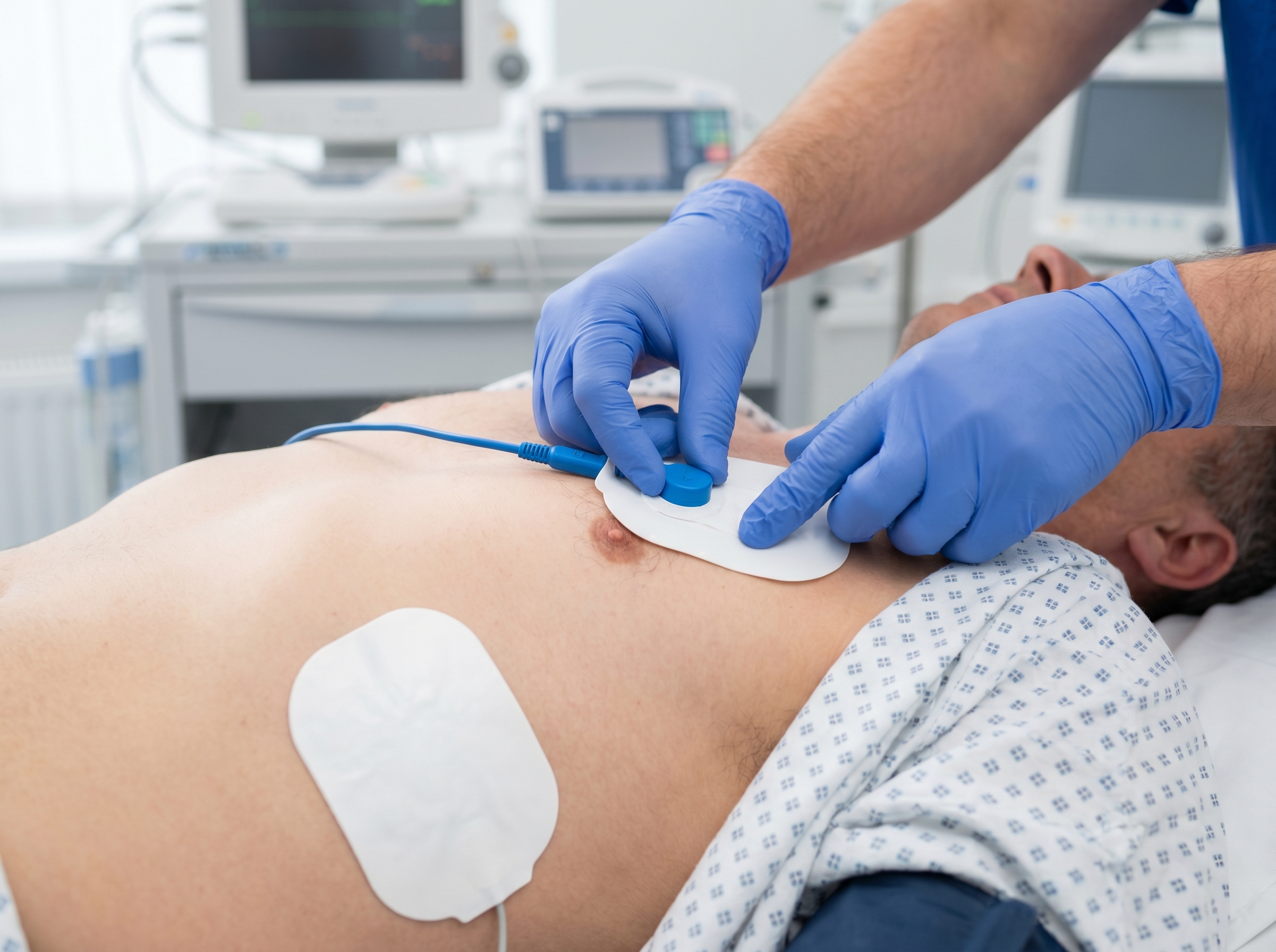
The anterolateral (AL) position is the default for most defibrillation scenarios. The sternal (right) pad is placed on the right side of the sternum, just below the right clavicle. The apical (left) pad is placed at the left mid-axillary line, over the apex of the heart, approximately at the V6 ECG position. This placement creates a vector that passes through the bulk of the left ventricle — the target myocardium for VF termination.
For AEDs, the anterolateral position is what the diagram on the pads instructs. Lay rescuers should follow this guidance without modification. For patients with implanted cardiac devices like pacemakers or ICDs, pad placement needs a minor modification: position pads at least one inch (2.5 cm) away from the device generator to avoid current shunting through the lead wires, which can both reduce defibrillation efficacy and damage the device.
The anteroposterior (AP) pad position places the anterior pad on the left precordium — roughly at the V2 to V4 ECG position — and the posterior pad on the back, left of the spine, below the left scapula. This creates a current vector that passes directly through the myocardium from front to back.
A systematic review published in the NIH PMC database on pad placement and return of spontaneous circulation found that anteroposterior placement may be associated with higher rates of VF termination. One cluster randomized trial reported termination rates of 79.9% with AP positioning versus 67.6% with anterolateral placement. The 2025 AHA guidelines acknowledge AP placement as an acceptable alternative, and emerging evidence suggests it may be preferred in refractory cases or when initial anterolateral shocks fail.
In practice, AP placement is logistically challenging during active CPR — rolling a patient to place a posterior pad interrupts compressions. The anterolateral position remains first-line for most arrest scenarios. However, if initial shocks fail and you are reassessing your resuscitation approach, considering a pad position change to AP is a clinically reasonable escalation step alongside antiarrhythmic escalation.
The biaxillary position — both pads in the mid-axillary line, one on each side — is another validated alternative, particularly useful in patients with central chest devices, dressings, or anatomic constraints that prevent standard anterolateral placement. ILCOR guidance confirms that anterolateral, anteroposterior, and biaxillary positions are all clinically acceptable in adults, with no single position definitively proven superior in all scenarios. The key principle: maximize current density through the myocardium while avoiding impedance-increasing obstacles such as pacemaker generators, sternotomy wires, or transcutaneous patches.
Even when energy settings and pad position are optimal, transthoracic impedance can significantly reduce the current that actually reaches the myocardium. Impedance is affected by pad-to-skin contact quality, chest hair, body habitus, lung inflation state, and pad electrode gel condition. Understanding impedance helps you troubleshoot when initial shocks fail.
Modern biphasic defibrillators measure transthoracic impedance before each shock and adjust waveform characteristics in real time to compensate. This is one of the primary reasons biphasic waveforms outperform monophasic devices — the adaptive compensation ensures consistent transmyocardial current density regardless of patient-specific impedance variables. A comprehensive StatPearls review of defibrillation on NCBI Bookshelf covers the electrical physiology of impedance compensation in depth for providers who want to go further on this topic.
The relationship between CPR and shock delivery is one of the most evidence-rich areas of resuscitation science, and the current recommendations are clear: minimize pre-shock pauses, resume CPR immediately after every shock, and do not check the rhythm immediately after a shock.
High-quality CPR generates forward coronary perfusion pressure — the physiologic condition that makes the myocardium receptive to defibrillation. When compressions stop, coronary perfusion pressure falls within seconds. A pre-shock pause (the time from last compression to shock delivery) of more than five seconds significantly reduces first-shock success rates. AHA guidelines currently target a pre-shock pause of less than five seconds.
In practice, this means charging the defibrillator while CPR is still in progress, completing the safety sweep while the team continues compressions, and delivering the shock within seconds of the last compression. Training teams to execute this choreography precisely is a core function of effective ACLS team dynamics — the team leader calls the charge, compressions continue, and everyone is positioned for a near-seamless transition to shock delivery and immediate CPR resumption.
One of the most important behavioral shifts from older ACLS protocols is the elimination of the immediate post-shock rhythm check. Current guidelines mandate that CPR resume immediately after shock delivery — without pausing to check the rhythm. The reason is physiologic: even when defibrillation successfully terminates VF, the myocardium requires several seconds to organize a perfusing rhythm. Immediately checking the pulse or rhythm wastes exactly the time when the heart needs coronary perfusion support most.
After two minutes of post-shock CPR, the team pauses for a rhythm check. If the rhythm has organized, check for a pulse. If VF has returned or persisted, prepare to shock again. This 2-minute cycle structure is the foundation of the ACLS cardiac arrest algorithm. ACLS medication timing is integrated into this same 2-minute cycle — epinephrine is given every 3 to 5 minutes, and antiarrhythmics like amiodarone or lidocaine are administered after the third shock in refractory VF.
When VF persists after three or more shocks despite high-quality CPR and appropriate medication delivery, the resuscitation team faces a genuinely difficult clinical problem. Several escalation strategies are supported by current evidence.
Double sequential defibrillation (DSD) — delivering two near-simultaneous shocks from two defibrillators placed with pads in different vectors — has gained clinical interest as a strategy for shock-refractory VF. Some case series and small trials show improved termination rates. While not yet a formal first-line ACLS recommendation, DSD is performed at many high-volume resuscitation centers as a rescue strategy and remains an active area of guideline discussion.
Antiarrhythmic selection matters in refractory VF as well. The choice between lidocaine and amiodarone for shock-refractory VF and VT has meaningful clinical implications — both are endorsed by current ACLS guidelines, but their pharmacologic profiles differ in ways that can affect which is optimal in specific patient contexts. After antiarrhythmic delivery, reassessing pad position as an additional escalation option is clinically supported by the evidence on AP placement.
Correcting reversible causes — the H's and T's — is always concurrent with defibrillation attempts. Hypokalemia, hypomagnesemia, and hypothermia all increase defibrillation resistance and should be actively investigated and treated in refractory cases. Understanding the reversible causes of pulseless rhythms is foundational ACLS knowledge that directly informs how you manage a refractory arrest, because an untreated cause will defeat every shock you deliver.
Standard defibrillation protocols cover the majority of cardiac arrest cases, but several patient populations require modified thinking on energy, pad placement, or procedural steps.
Defibrillation is not contraindicated in pregnancy — the energy delivered to the fetus through amniotic fluid is minimal, and the maternal arrhythmia absolutely must be treated. Use standard energy settings and pad positions without modification for the rhythm type. Avoid placing pads over the uterine fundus when possible, but do not delay treatment to achieve perfect positioning. Left lateral uterine displacement should be maintained by a dedicated team member throughout resuscitation to relieve aortocaval compression, which otherwise severely limits CPR efficacy.
Pediatric defibrillation follows weight-based dosing. The initial dose is 2 J/kg for VF and pulseless VT, escalating to 4 J/kg for subsequent shocks. Many AEDs now include pediatric pads or a pediatric mode that automatically applies dose attenuation for children under eight years or under 25 kilograms. Manual pediatric defibrillation requires provider calculation — knowing that a 20 kg child receives a 40 J initial shock and an 80 J escalation dose. According to the StatPearls guide to synchronized cardioversion on NCBI Bookshelf, pediatric cardioversion dosing similarly follows weight-based scaling at 0.5 to 1 J/kg initial dose, escalating to 2 J/kg.
Obesity increases transthoracic impedance due to chest wall adipose tissue and may alter the geometry of pad position relative to the underlying heart. Standard energy settings remain appropriate — do not reduce initial energy doses in obese patients; if anything, the higher baseline impedance argues for ensuring you are not under-delivering. Check pad placement carefully to ensure the full electrode surface is adhering to the chest wall, particularly in the apical position on a large chest. The biaxillary pad position can be a useful alternative when anterolateral placement is constrained by body habitus or by pannus anatomy limiting pad adhesion.
Understanding the clinical science of defibrillation is essential, but operationalizing it under pressure requires systematic training. ACLS certification teaches providers not just what to do, but how to execute interventions with the efficiency and precision that saves lives in real codes.
Affordable ACLS offers physician-developed ACLS certification and recertification courses built around current AHA and ILCOR guidelines — including the updated 2025 recommendations on defibrillation energy dosing, pad placement, and shock timing. Courses are fully online, self-paced, and typically completed in one to two hours, making them genuinely accessible for working clinicians who cannot afford to block an entire day for a hospital-based skills session.
The curriculum covers the complete cardiac arrest algorithm: shockable versus non-shockable rhythms, defibrillation technique, synchronized cardioversion, airway management, ACLS pharmacology, and post-resuscitation care. Every module reflects what practicing emergency physicians apply clinically — because Affordable ACLS was built by Board Certified Emergency Medicine physicians with 20-plus years of combined clinical and academic experience. ACLS Certification is available for $99, with recertification at $89, and all certifications include unlimited retakes at no extra charge plus a money-back guarantee if your employer does not accept the credential.
For providers who need both ACLS and BLS, bundles start at $123 — making comprehensive certification achievable without the financial barrier that often delays compliance. If you are ready to build confidence in every component of cardiac arrest management, including the defibrillation decisions covered in this article, explore the difference between BLS and ACLS certification to confirm which credential fits your practice setting and scope.
Manual and automated defibrillation both serve critical roles in the cardiac arrest response ecosystem, but they demand different levels of clinical knowledge from the providers using them. AEDs empower lay rescuers to act decisively before trained help arrives. Manual defibrillators give ACLS providers the full range of electrical therapy options — with all the clinical judgment that comes with them.
The key takeaways from this deep dive: use biphasic energy whenever possible and know your device's recommended dose; match energy settings to the specific arrhythmia and always synchronize when cardioverting a patient with a pulse; understand the anterior-lateral position as your default and the anteroposterior position as a viable escalation for refractory VF; minimize every pre- and post-shock pause to preserve the coronary perfusion pressure your CPR has built; and escalate systematically through antiarrhythmics, pad position changes, and reversible cause correction when initial shocks fail.
These are not abstract principles — they are the exact decisions you will face the next time a patient's rhythm demands electrical therapy. For providers who want to sharpen all of these skills in a structured, guideline-compliant framework, ACLS certification through Affordable ACLS provides the clinical depth, flexibility, and credentialing you need, on your schedule, at a price that reflects what working healthcare providers actually deserve.
.jpg)