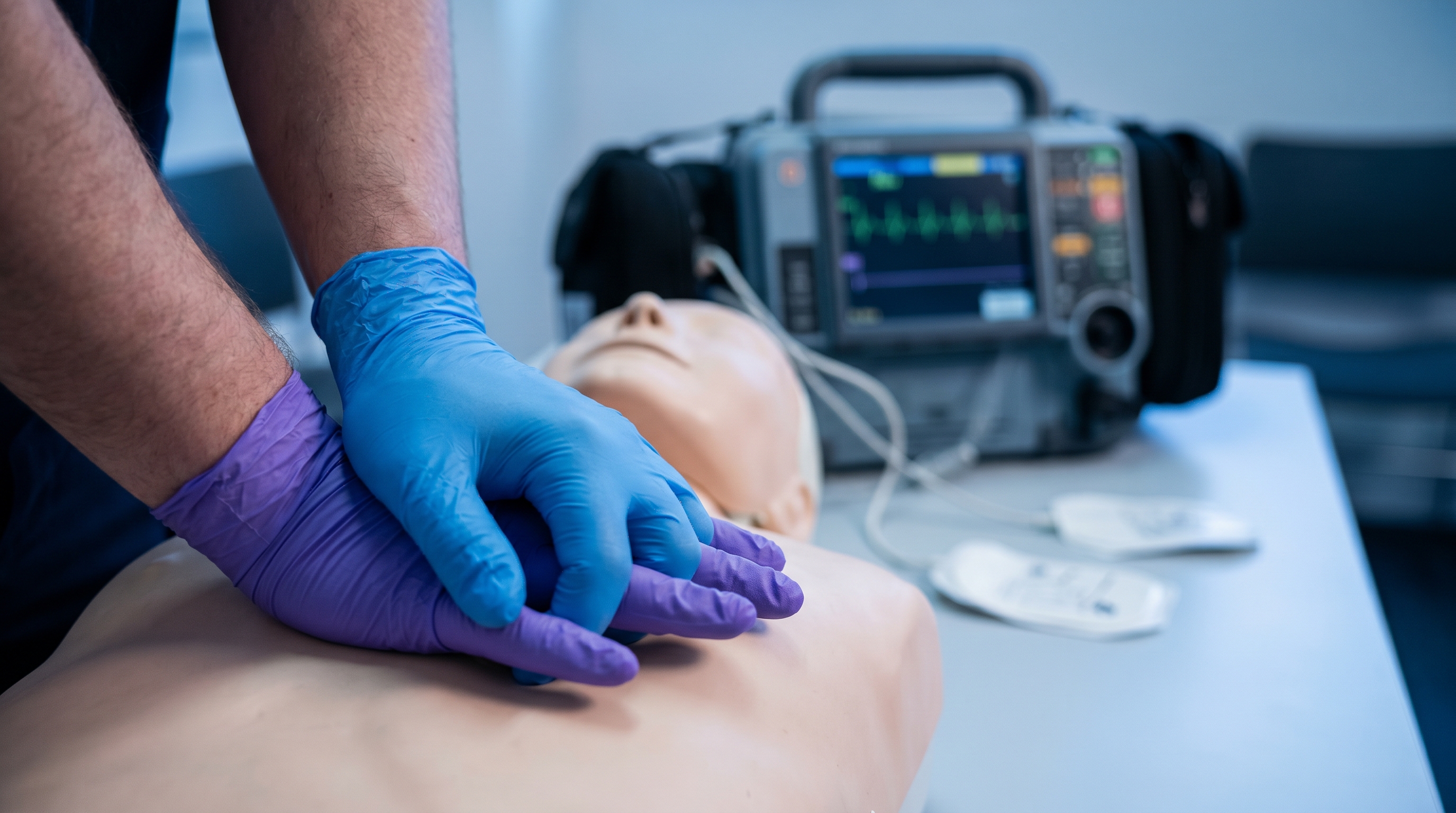
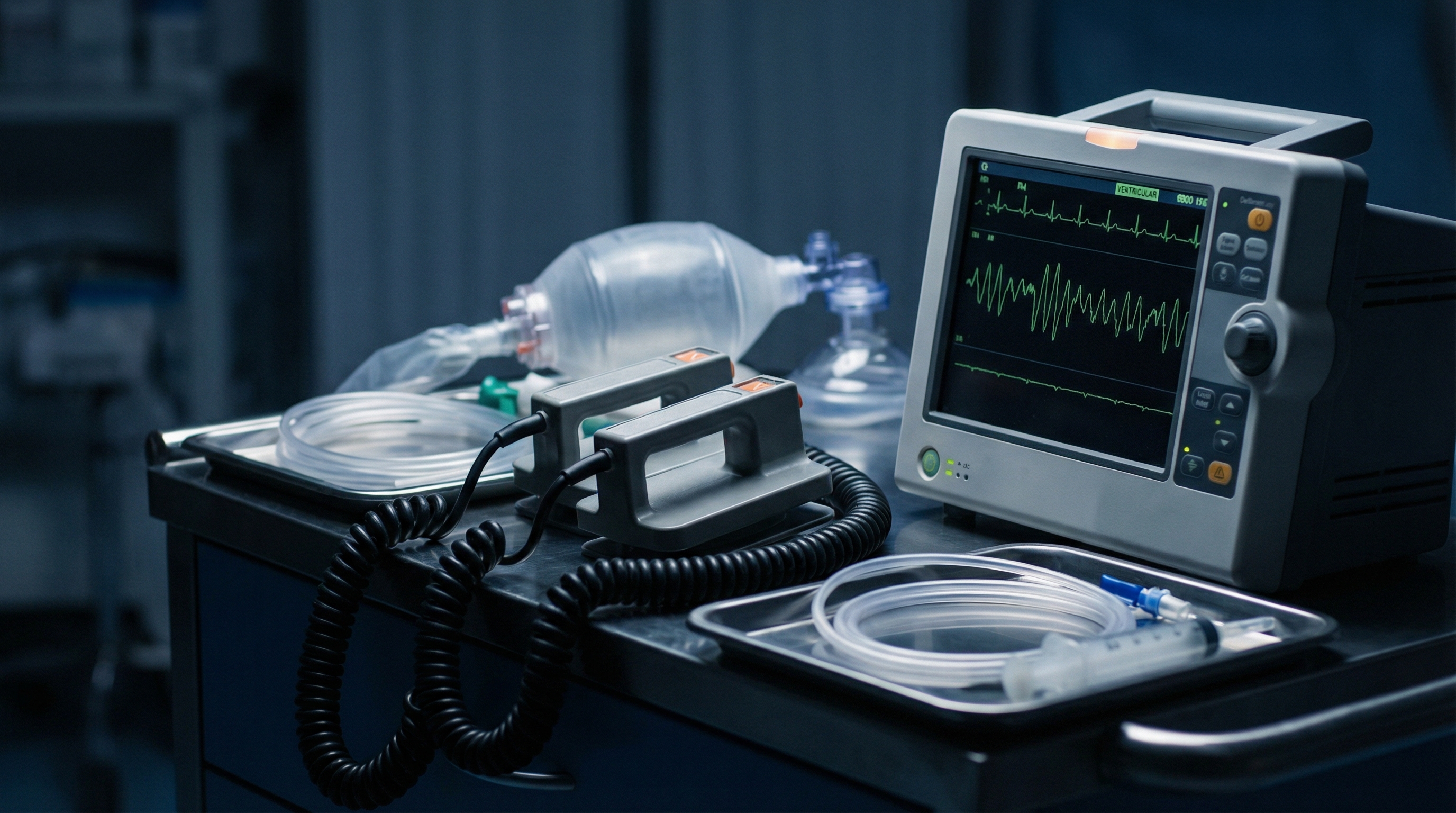
ACLS Blogs

ACLS Megacode Scenarios: 5 Full Practice Cases to Run Mentally

Megacode day tests composure as much as it tests knowledge. On the actual station, you do not get a printed algorithm to follow along with - you get a monitor, a mannequin, and an evaluator watching you make calls in real time. One of the cheapest ways to build that composure before test day costs nothing and needs no equipment: run the case in your head. Close your eyes, picture the monitor, and walk yourself through the decision at each fork in the algorithm, out loud if you can manage it. This article gives you five complete megacode cases, each self-contained, that you can run mentally anywhere - in your car before a shift, on a break, or the night before your exam.


This is Volume 1 of a scenario series. If you have not already worked through how the actual test stations flow, start with the station-by-station megacode walkthrough for context on how a live megacode is structured before dropping into these five cases. Each case below gives you the presentation, the decision points where candidates typically freeze, the correct actions with the reasoning behind them, and the specific errors that cost points on a real station.


How to Run These Scenarios in Your Head

Read the presentation for a case, then stop. Before reading any further, say out loud, or write down, what you would do first, second, and third. Only after you have committed to an answer should you read the decision points and correct actions to check your reasoning. This forces you to actually decide the way you will have to on test day, instead of passively recognizing the right answer once it is already sitting in front of you.


Pair this drill with a timed check on recall, since running scenarios mentally tests judgment but not speed. Our free practice tests are useful for that second half of the gap check right after you talk through a case. If you are working through your last week of prep, the 1-week ACLS study plan for busy clinicians shows where scenario drills like these fit alongside the rest of your review.


Case 1: Witnessed Ventricular Fibrillation Arrest

You are standing at the nurses station watching a telemetry screen when a patient's monitor alarms. You look up as the rhythm changes to a coarse, chaotic pattern with no organized QRS complexes. You are at the bedside within seconds. The patient is unresponsive, not breathing normally, and has no palpable pulse.
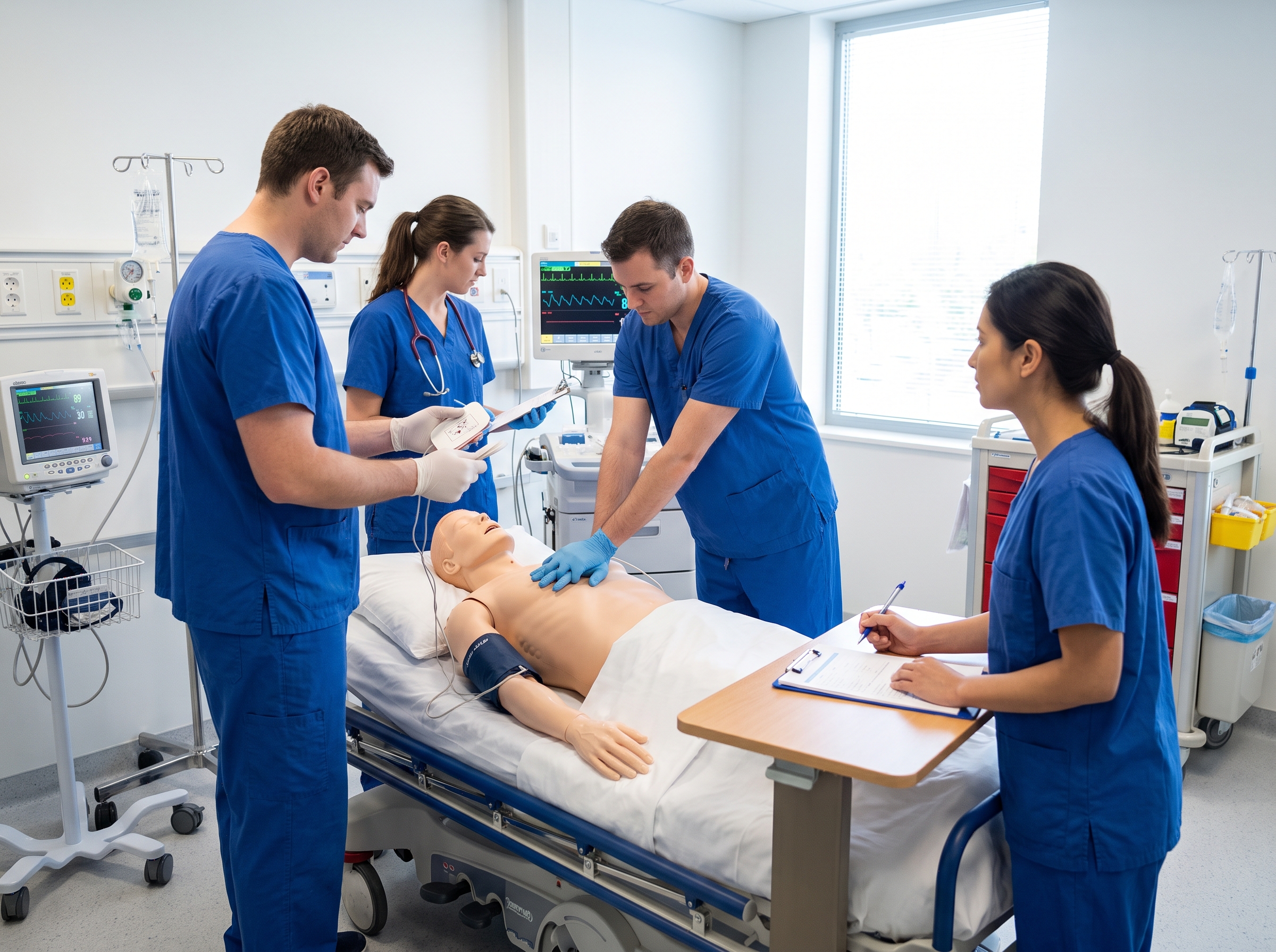
Emergency team practicing defibrillation and chest compressions on a code mannequin during ACLS training


Decision Points

Because this arrest is witnessed and monitored, the temptation is to freeze while someone finds the defibrillator. The decision that matters most in the first thirty seconds is simple: start compressions immediately and keep them going while pads are placed and the device charges. The second decision point is what happens the instant after the first shock - do you pause to check a pulse, or resume compressions right away? The third is when drugs enter the sequence and in what order.


Correct Actions and Rationale

Start high-quality chest compressions immediately at a rate of 100 to 120 per minute, at least 2 inches deep, allowing full chest recoil between compressions, while a second team member gets pads on the patient. As soon as the defibrillator is charged and ready, deliver one shock at the energy your specific device recommends, then resume compressions immediately for a full 2-minute cycle. Do not stop to feel for a pulse right after the shock - the rhythm is still very likely to be disorganized, and every second off the chest lowers coronary perfusion pressure.


Get IV or IO access during compressions, not instead of them. If the patient is still in ventricular fibrillation or pulseless ventricular tachycardia after the second shock, give epinephrine 1 mg IV or IO and repeat this dose every 3 to 5 minutes for the remainder of the arrest, per the AHA Adult Cardiac Arrest Algorithm. If the shockable rhythm persists after the third shock, give amiodarone 300 mg IV or IO as a bolus; if it recurs again, a second dose of 150 mg IV or IO is appropriate. Only recheck the rhythm at the end of each 2-minute cycle, and only check for a pulse if that rhythm looks organized.


Rotate the compressor every 2-minute cycle even if the current compressor feels fine, since compression quality reliably decays with fatigue well before a person notices it. Being able to identify an organized rhythm quickly at each check matters here - a fast rhythm recognition reference is worth having memorized cold before you ever reach this station.


Common Errors That Cost Points

  • Stopping compressions for a pulse check immediately after defibrillation instead of resuming compressions right away.
  • Pushing epinephrine and amiodarone back to back through the same line without a flush, or pausing compressions to give a drug.
  • Letting compression depth and rate drift down over the last few cycles without rotating the compressor.
  • Losing track of the shock count and drug timing under stress instead of verbalizing the cycle count and clock out loud.

Case 2: PEA Arrest With a Reversible Cause to Find

A post-operative patient becomes unresponsive. The monitor shows a narrow, organized rhythm at 70 beats per minute, but you cannot feel a pulse and the patient is not breathing normally. A family member mentions the patient has had new shortness of breath and one-sided chest discomfort since a central line was placed earlier that day.


Decision Points

The first trap is letting an organized rhythm on the monitor talk you out of believing there is truly no pulse. The second is defaulting to "shock the rhythm" out of habit from a VF case - this is pulseless electrical activity, and it is not a shockable rhythm. The third, and the one the stem is built around, is whether you work through the reversible causes systematically or simply push epinephrine on a loop and hope.


Correct Actions and Rationale

Begin CPR immediately using the same quality standards as any arrest, and get IV or IO access. Give epinephrine 1 mg IV or IO every 3 to 5 minutes; because PEA is a non-shockable rhythm, there is no role for a defibrillator here. Between epinephrine doses, work through the reversible causes rather than repeating the drug alone: hypovolemia, hypoxia, hydrogen ion excess (acidosis), hypo- or hyperkalemia, hypothermia, tension pneumothorax, cardiac tamponade, toxins, and thrombosis, whether pulmonary or coronary. According to the NIH StatPearls review of pulseless electrical activity, outcomes only improve meaningfully when a reversible cause is found and corrected quickly, which is exactly what this station is testing.


  • Hypovolemia
  • Hypoxia
  • Hydrogen ion (acidosis)
  • Hypo- or hyperkalemia
  • Hypothermia
  • Tension pneumothorax
  • Cardiac tamponade
  • Toxins
  • Thrombosis, pulmonary or coronary

The case clue here (a fresh central line, one-sided chest discomfort, new shortness of breath) is pointing you toward tension pneumothorax from the procedure, which needs needle decompression rather than another round of epinephrine alone. Holding a nine-item differential under pressure is genuinely hard, so it helps to drill it the same way you drill drug doses - the algorithm memory hacks and mnemonics guide covers a few ways to keep this list retrievable when you are also managing compressions, a clock, and a chart.


Common Errors That Cost Points

  • Treating PEA as if there is nothing to do but push epinephrine, and never systematically working the differential.
  • Attempting to defibrillate an organized PEA rhythm out of habit from a shockable-rhythm case.
  • Missing a context clue in the stem, such as a recent procedure or one-sided symptoms, that examiners plant specifically to point toward one reversible cause.
  • Delaying a bedside intervention like needle decompression while waiting for imaging that will never be available during a code.

Case 3: Asystole and the Decision to Stop

A nursing assistant finds a patient unresponsive on the floor. The time down is unknown, and the arrest was not witnessed. CPR is already in progress by the time you arrive as the code leader, and the monitor shows a flat line.


Decision Points

The first decision is whether the flat line is really asystole, or an equipment problem - a loose lead or a low gain setting can mimic it. The second is recognizing that, like PEA, asystole is a non-shockable rhythm, so there is no defibrillation step to reach for. The hardest decision in this case is not clinical at all: it is when, and how, to raise the question of stopping resuscitation.


Correct Actions and Rationale

Confirm the flat line in more than one lead and check the equipment (leads, gain, connections) before accepting asystole as real; a disconnected lead is a distractor examiners use deliberately. Continue high-quality CPR, secure IV or IO access, and give epinephrine 1 mg IV or IO every 3 to 5 minutes. Search for and treat reversible causes the same way you would in PEA, since an unwitnessed arrest with an unknown down time raises the index of suspicion for hypoxia, hypovolemia, hypothermia, or a metabolic cause. Reviewing the full branch-by-branch arrest algorithm walkthrough makes it clear that asystole and PEA share the same drug and reversible-cause pathway, and only split off from ventricular fibrillation and pulseless ventricular tachycardia at the shock decision.


Current AHA guidance does not set a fixed time limit for when to stop resuscitation; the decision weighs whether the arrest was witnessed and monitored, the initial rhythm, how long resuscitation has continued without any organized rhythm or return of spontaneous circulation, and whether reversible causes have genuinely been excluded. Research on termination-of-resuscitation decision rules for in-hospital cardiac arrest gives a sense of the factors clinicians weigh in that judgment call, rather than a script to recite. On a megacode station, verbalize this reasoning to your evaluator - stating why you are considering stopping, such as an unwitnessed arrest with prolonged asystole despite adequate CPR, appropriate epinephrine dosing, and no reversible cause found, is itself part of what is being assessed.


Common Errors That Cost Points

  • Shocking asystole because a candidate defaults to shock-first reflexes carried over from the VF case.
  • Failing to confirm the rhythm in a second lead and missing a lead-disconnect distractor built into the scenario.
  • Either stopping without ever verbalizing the reasoning, or never raising the question at all, both of which read as a candidate who has not thought the decision through.
  • Giving amiodarone out of habit, even though it has no role in a non-shockable rhythm.

Case 4: Unstable Bradycardia Progressing to Pacing

A patient on telemetry develops a heart rate of 38 beats per minute. The patient is diaphoretic, confused, and has a blood pressure of 78/50. Because there is a pulse and the patient is breathing, this is not a cardiac arrest, but something is clearly wrong and getting worse.
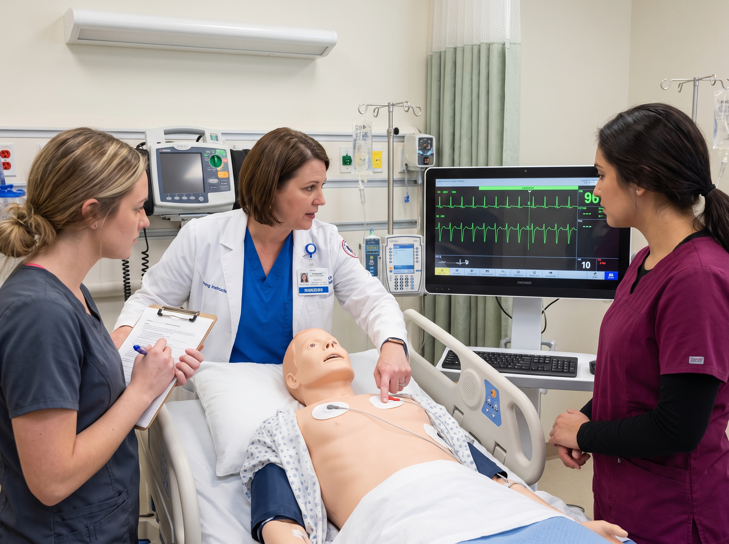
Instructor demonstrating transcutaneous pacing pad placement during ACLS bradycardia training


Decision Points

The first decision is confirming that this bradycardia is symptomatic and unstable, based on hypotension and altered mental status, rather than an incidental slow rate that needs no acute treatment. The second is knowing the ceiling on the first-line drug before you get there. The third, and the one that separates a strong candidate from a weak one, is recognizing when to stop waiting on medication and escalate to pacing.


Correct Actions and Rationale

Confirm the patient is symptomatic and unstable, place them on the monitor, get IV access, and prepare for the possibility of pacing early rather than only after several failed medication attempts. Give atropine 1 mg IV, and repeat this dose every 3 to 5 minutes to a maximum total dose of 3 mg, per the AHA Adult Bradycardia With a Pulse Algorithm. If atropine is ineffective, or the block looks unlikely to respond to it, such as a high-degree AV block, move to transcutaneous pacing without waiting on further doses, or consider a dopamine or epinephrine infusion as an alternative if pacing is not immediately available.


If you initiate pacing, verbalize confirming mechanical capture, not just electrical capture. Spikes on the monitor that line up with the paced rate are not enough on their own; you need a palpable pulse that matches that rate before you call it successful.


Common Errors That Cost Points

  • Redosing atropine repeatedly past the point of clinical response while the patient continues to deteriorate, instead of escalating to pacing.
  • Confusing electrical capture on the monitor with mechanical capture, and declaring success before confirming a pulse.
  • Treating a stable, asymptomatic bradycardia with the same urgency as this unstable case.
  • Forgetting that atropine has a ceiling dose and continuing to redose past 3 mg total.

Case 5: Unstable Tachycardia Requiring Synchronized Cardioversion

A patient's monitor alarms for a heart rate of 172. The rhythm is narrow-complex and regular. The patient is diaphoretic, lightheaded, has ongoing chest discomfort, and a blood pressure of 82/54.


Decision Points

The first decision is stable versus unstable - this patient's hypotension and symptoms make this case unstable, which changes the entire pathway compared with a stable, narrow-complex tachycardia you might manage with vagal maneuvers or adenosine first. The second is choosing synchronized cardioversion over defibrillation, and understanding why synchronization matters at all. The third is recognizing what changes if the rhythm were irregular or wide-complex instead of the regular narrow-complex rhythm actually presented here.


Correct Actions and Rationale

Because the patient is unstable but still has a pulse, this calls for synchronized cardioversion, not defibrillation. Synchronization times the shock to fire on the R wave so it does not land on the T wave and provoke ventricular fibrillation. If equipment and the patient's condition allow without meaningfully delaying care, brief sedation is appropriate before cardioversion in a real patient; on a megacode station, you will typically just state that you would sedate rather than perform it.


Select the energy your defibrillator's device-specific settings recommend for the rhythm. Current AHA guidance has moved away from prescribing one fixed number for every device and instead directs providers to the manufacturer's recommended setting for the rhythm being treated, as reflected in the AHA Electrical Cardioversion Algorithm. Many courses still teach a practical starting range of roughly 100 J biphasic for a regular narrow-complex rhythm like this one, escalating toward 120 to 200 J biphasic for a rhythm such as atrial fibrillation if the first shock does not convert it. If the rhythm were instead irregularly irregular and wide-complex, such as polymorphic ventricular tachycardia, synchronization may not fire reliably, and you would treat it as you would treat ventricular fibrillation, with an unsynchronized, high-energy shock. Recognizing that branch point on the monitor is often the real test.


Reassess the rhythm and pulse immediately after cardioversion, and be ready to escalate energy or add an antiarrhythmic if the first shock does not convert the rhythm.


Common Errors That Cost Points

  • Defibrillating, unsynchronized, an unstable but pulsed tachycardia, risking a shock landing on a T wave.
  • Forgetting that most defibrillators default back to unsynchronized mode after every delivered shock, so sync has to be manually reselected before a second cardioversion attempt.
  • Misidentifying an irregular or wide-complex rhythm as the straightforward regular, narrow-complex case and choosing the wrong energy pathway.
  • Delaying cardioversion to give repeated doses of a rate-control drug to a patient who is already unstable.

Keep the Reps Going

These five cases cover the branch points examiners return to again and again: the shockable-versus-non-shockable fork in cardiac arrest, the reversible-cause search that separates a strong PEA or asystole response from a weak one, the decision to escalate a symptomatic bradycardia to pacing instead of redosing medication indefinitely, and the synchronized-versus-unsynchronized choice in an unstable tachycardia. Running them mentally and out loud on a regular basis builds the kind of automatic recall you need when an evaluator is standing over your shoulder with a stopwatch.


  • Case 1: Witnessed VF arrest - compressions first, shock, then resume compressions immediately, epinephrine after the second shock, amiodarone after the third.
  • Case 2: PEA arrest - no shock, epinephrine on schedule, and a systematic search through the reversible causes.
  • Case 3: Asystole - confirm the rhythm in a second lead, no shock, same reversible-cause search, and a verbalized decision about stopping.
  • Case 4: Unstable bradycardia - atropine to a 3 mg ceiling, then pacing or an infusion, with mechanical capture confirmed by pulse.
  • Case 5: Unstable tachycardia - synchronized cardioversion at a device-appropriate energy, re-synced before any repeat shock.

Once these five feel automatic, test that recall under time pressure with the ACLS pretest of 20 practice questions with answer rationales, and watch for Volume 2 of this series for five more cases built around branch points these did not cover.


Mental reps build the confidence to get through the room, but the credential itself is what your employer or state board actually requires. Whether you are certifying for the first time or your card has lapsed while you were focused on studying, you can complete the full ACLS certification course online at your own pace and put these five scenarios to work on your actual exam.


ACLS Blogs

ACLS Megacode Scenarios: 5 Full Practice Cases to Run Mentally

Megacode day tests composure as much as it tests knowledge. On the actual station, you do not get a printed algorithm to follow along with - you get a monitor, a mannequin, and an evaluator watching you make calls in real time. One of the cheapest ways to build that composure before test day costs nothing and needs no equipment: run the case in your head. Close your eyes, picture the monitor, and walk yourself through the decision at each fork in the algorithm, out loud if you can manage it. This article gives you five complete megacode cases, each self-contained, that you can run mentally anywhere - in your car before a shift, on a break, or the night before your exam.


This is Volume 1 of a scenario series. If you have not already worked through how the actual test stations flow, start with the station-by-station megacode walkthrough for context on how a live megacode is structured before dropping into these five cases. Each case below gives you the presentation, the decision points where candidates typically freeze, the correct actions with the reasoning behind them, and the specific errors that cost points on a real station.


How to Run These Scenarios in Your Head

Read the presentation for a case, then stop. Before reading any further, say out loud, or write down, what you would do first, second, and third. Only after you have committed to an answer should you read the decision points and correct actions to check your reasoning. This forces you to actually decide the way you will have to on test day, instead of passively recognizing the right answer once it is already sitting in front of you.


Pair this drill with a timed check on recall, since running scenarios mentally tests judgment but not speed. Our free practice tests are useful for that second half of the gap check right after you talk through a case. If you are working through your last week of prep, the 1-week ACLS study plan for busy clinicians shows where scenario drills like these fit alongside the rest of your review.


Case 1: Witnessed Ventricular Fibrillation Arrest

You are standing at the nurses station watching a telemetry screen when a patient's monitor alarms. You look up as the rhythm changes to a coarse, chaotic pattern with no organized QRS complexes. You are at the bedside within seconds. The patient is unresponsive, not breathing normally, and has no palpable pulse.

Emergency team practicing defibrillation and chest compressions on a code mannequin during ACLS training


Decision Points

Because this arrest is witnessed and monitored, the temptation is to freeze while someone finds the defibrillator. The decision that matters most in the first thirty seconds is simple: start compressions immediately and keep them going while pads are placed and the device charges. The second decision point is what happens the instant after the first shock - do you pause to check a pulse, or resume compressions right away? The third is when drugs enter the sequence and in what order.


Correct Actions and Rationale

Start high-quality chest compressions immediately at a rate of 100 to 120 per minute, at least 2 inches deep, allowing full chest recoil between compressions, while a second team member gets pads on the patient. As soon as the defibrillator is charged and ready, deliver one shock at the energy your specific device recommends, then resume compressions immediately for a full 2-minute cycle. Do not stop to feel for a pulse right after the shock - the rhythm is still very likely to be disorganized, and every second off the chest lowers coronary perfusion pressure.


Get IV or IO access during compressions, not instead of them. If the patient is still in ventricular fibrillation or pulseless ventricular tachycardia after the second shock, give epinephrine 1 mg IV or IO and repeat this dose every 3 to 5 minutes for the remainder of the arrest, per the AHA Adult Cardiac Arrest Algorithm. If the shockable rhythm persists after the third shock, give amiodarone 300 mg IV or IO as a bolus; if it recurs again, a second dose of 150 mg IV or IO is appropriate. Only recheck the rhythm at the end of each 2-minute cycle, and only check for a pulse if that rhythm looks organized.


Rotate the compressor every 2-minute cycle even if the current compressor feels fine, since compression quality reliably decays with fatigue well before a person notices it. Being able to identify an organized rhythm quickly at each check matters here - a fast rhythm recognition reference is worth having memorized cold before you ever reach this station.


Common Errors That Cost Points

  • Stopping compressions for a pulse check immediately after defibrillation instead of resuming compressions right away.
  • Pushing epinephrine and amiodarone back to back through the same line without a flush, or pausing compressions to give a drug.
  • Letting compression depth and rate drift down over the last few cycles without rotating the compressor.
  • Losing track of the shock count and drug timing under stress instead of verbalizing the cycle count and clock out loud.

Case 2: PEA Arrest With a Reversible Cause to Find

A post-operative patient becomes unresponsive. The monitor shows a narrow, organized rhythm at 70 beats per minute, but you cannot feel a pulse and the patient is not breathing normally. A family member mentions the patient has had new shortness of breath and one-sided chest discomfort since a central line was placed earlier that day.


Decision Points

The first trap is letting an organized rhythm on the monitor talk you out of believing there is truly no pulse. The second is defaulting to "shock the rhythm" out of habit from a VF case - this is pulseless electrical activity, and it is not a shockable rhythm. The third, and the one the stem is built around, is whether you work through the reversible causes systematically or simply push epinephrine on a loop and hope.


Correct Actions and Rationale

Begin CPR immediately using the same quality standards as any arrest, and get IV or IO access. Give epinephrine 1 mg IV or IO every 3 to 5 minutes; because PEA is a non-shockable rhythm, there is no role for a defibrillator here. Between epinephrine doses, work through the reversible causes rather than repeating the drug alone: hypovolemia, hypoxia, hydrogen ion excess (acidosis), hypo- or hyperkalemia, hypothermia, tension pneumothorax, cardiac tamponade, toxins, and thrombosis, whether pulmonary or coronary. According to the NIH StatPearls review of pulseless electrical activity, outcomes only improve meaningfully when a reversible cause is found and corrected quickly, which is exactly what this station is testing.


  • Hypovolemia
  • Hypoxia
  • Hydrogen ion (acidosis)
  • Hypo- or hyperkalemia
  • Hypothermia
  • Tension pneumothorax
  • Cardiac tamponade
  • Toxins
  • Thrombosis, pulmonary or coronary

The case clue here (a fresh central line, one-sided chest discomfort, new shortness of breath) is pointing you toward tension pneumothorax from the procedure, which needs needle decompression rather than another round of epinephrine alone. Holding a nine-item differential under pressure is genuinely hard, so it helps to drill it the same way you drill drug doses - the algorithm memory hacks and mnemonics guide covers a few ways to keep this list retrievable when you are also managing compressions, a clock, and a chart.


Common Errors That Cost Points

  • Treating PEA as if there is nothing to do but push epinephrine, and never systematically working the differential.
  • Attempting to defibrillate an organized PEA rhythm out of habit from a shockable-rhythm case.
  • Missing a context clue in the stem, such as a recent procedure or one-sided symptoms, that examiners plant specifically to point toward one reversible cause.
  • Delaying a bedside intervention like needle decompression while waiting for imaging that will never be available during a code.

Case 3: Asystole and the Decision to Stop

A nursing assistant finds a patient unresponsive on the floor. The time down is unknown, and the arrest was not witnessed. CPR is already in progress by the time you arrive as the code leader, and the monitor shows a flat line.


Decision Points

The first decision is whether the flat line is really asystole, or an equipment problem - a loose lead or a low gain setting can mimic it. The second is recognizing that, like PEA, asystole is a non-shockable rhythm, so there is no defibrillation step to reach for. The hardest decision in this case is not clinical at all: it is when, and how, to raise the question of stopping resuscitation.


Correct Actions and Rationale

Confirm the flat line in more than one lead and check the equipment (leads, gain, connections) before accepting asystole as real; a disconnected lead is a distractor examiners use deliberately. Continue high-quality CPR, secure IV or IO access, and give epinephrine 1 mg IV or IO every 3 to 5 minutes. Search for and treat reversible causes the same way you would in PEA, since an unwitnessed arrest with an unknown down time raises the index of suspicion for hypoxia, hypovolemia, hypothermia, or a metabolic cause. Reviewing the full branch-by-branch arrest algorithm walkthrough makes it clear that asystole and PEA share the same drug and reversible-cause pathway, and only split off from ventricular fibrillation and pulseless ventricular tachycardia at the shock decision.


Current AHA guidance does not set a fixed time limit for when to stop resuscitation; the decision weighs whether the arrest was witnessed and monitored, the initial rhythm, how long resuscitation has continued without any organized rhythm or return of spontaneous circulation, and whether reversible causes have genuinely been excluded. Research on termination-of-resuscitation decision rules for in-hospital cardiac arrest gives a sense of the factors clinicians weigh in that judgment call, rather than a script to recite. On a megacode station, verbalize this reasoning to your evaluator - stating why you are considering stopping, such as an unwitnessed arrest with prolonged asystole despite adequate CPR, appropriate epinephrine dosing, and no reversible cause found, is itself part of what is being assessed.


Common Errors That Cost Points

  • Shocking asystole because a candidate defaults to shock-first reflexes carried over from the VF case.
  • Failing to confirm the rhythm in a second lead and missing a lead-disconnect distractor built into the scenario.
  • Either stopping without ever verbalizing the reasoning, or never raising the question at all, both of which read as a candidate who has not thought the decision through.
  • Giving amiodarone out of habit, even though it has no role in a non-shockable rhythm.

Case 4: Unstable Bradycardia Progressing to Pacing

A patient on telemetry develops a heart rate of 38 beats per minute. The patient is diaphoretic, confused, and has a blood pressure of 78/50. Because there is a pulse and the patient is breathing, this is not a cardiac arrest, but something is clearly wrong and getting worse.

Instructor demonstrating transcutaneous pacing pad placement during ACLS bradycardia training


Decision Points

The first decision is confirming that this bradycardia is symptomatic and unstable, based on hypotension and altered mental status, rather than an incidental slow rate that needs no acute treatment. The second is knowing the ceiling on the first-line drug before you get there. The third, and the one that separates a strong candidate from a weak one, is recognizing when to stop waiting on medication and escalate to pacing.


Correct Actions and Rationale

Confirm the patient is symptomatic and unstable, place them on the monitor, get IV access, and prepare for the possibility of pacing early rather than only after several failed medication attempts. Give atropine 1 mg IV, and repeat this dose every 3 to 5 minutes to a maximum total dose of 3 mg, per the AHA Adult Bradycardia With a Pulse Algorithm. If atropine is ineffective, or the block looks unlikely to respond to it, such as a high-degree AV block, move to transcutaneous pacing without waiting on further doses, or consider a dopamine or epinephrine infusion as an alternative if pacing is not immediately available.


If you initiate pacing, verbalize confirming mechanical capture, not just electrical capture. Spikes on the monitor that line up with the paced rate are not enough on their own; you need a palpable pulse that matches that rate before you call it successful.


Common Errors That Cost Points

  • Redosing atropine repeatedly past the point of clinical response while the patient continues to deteriorate, instead of escalating to pacing.
  • Confusing electrical capture on the monitor with mechanical capture, and declaring success before confirming a pulse.
  • Treating a stable, asymptomatic bradycardia with the same urgency as this unstable case.
  • Forgetting that atropine has a ceiling dose and continuing to redose past 3 mg total.

Case 5: Unstable Tachycardia Requiring Synchronized Cardioversion

A patient's monitor alarms for a heart rate of 172. The rhythm is narrow-complex and regular. The patient is diaphoretic, lightheaded, has ongoing chest discomfort, and a blood pressure of 82/54.


Decision Points

The first decision is stable versus unstable - this patient's hypotension and symptoms make this case unstable, which changes the entire pathway compared with a stable, narrow-complex tachycardia you might manage with vagal maneuvers or adenosine first. The second is choosing synchronized cardioversion over defibrillation, and understanding why synchronization matters at all. The third is recognizing what changes if the rhythm were irregular or wide-complex instead of the regular narrow-complex rhythm actually presented here.


Correct Actions and Rationale

Because the patient is unstable but still has a pulse, this calls for synchronized cardioversion, not defibrillation. Synchronization times the shock to fire on the R wave so it does not land on the T wave and provoke ventricular fibrillation. If equipment and the patient's condition allow without meaningfully delaying care, brief sedation is appropriate before cardioversion in a real patient; on a megacode station, you will typically just state that you would sedate rather than perform it.


Select the energy your defibrillator's device-specific settings recommend for the rhythm. Current AHA guidance has moved away from prescribing one fixed number for every device and instead directs providers to the manufacturer's recommended setting for the rhythm being treated, as reflected in the AHA Electrical Cardioversion Algorithm. Many courses still teach a practical starting range of roughly 100 J biphasic for a regular narrow-complex rhythm like this one, escalating toward 120 to 200 J biphasic for a rhythm such as atrial fibrillation if the first shock does not convert it. If the rhythm were instead irregularly irregular and wide-complex, such as polymorphic ventricular tachycardia, synchronization may not fire reliably, and you would treat it as you would treat ventricular fibrillation, with an unsynchronized, high-energy shock. Recognizing that branch point on the monitor is often the real test.


Reassess the rhythm and pulse immediately after cardioversion, and be ready to escalate energy or add an antiarrhythmic if the first shock does not convert the rhythm.


Common Errors That Cost Points

  • Defibrillating, unsynchronized, an unstable but pulsed tachycardia, risking a shock landing on a T wave.
  • Forgetting that most defibrillators default back to unsynchronized mode after every delivered shock, so sync has to be manually reselected before a second cardioversion attempt.
  • Misidentifying an irregular or wide-complex rhythm as the straightforward regular, narrow-complex case and choosing the wrong energy pathway.
  • Delaying cardioversion to give repeated doses of a rate-control drug to a patient who is already unstable.

Keep the Reps Going

These five cases cover the branch points examiners return to again and again: the shockable-versus-non-shockable fork in cardiac arrest, the reversible-cause search that separates a strong PEA or asystole response from a weak one, the decision to escalate a symptomatic bradycardia to pacing instead of redosing medication indefinitely, and the synchronized-versus-unsynchronized choice in an unstable tachycardia. Running them mentally and out loud on a regular basis builds the kind of automatic recall you need when an evaluator is standing over your shoulder with a stopwatch.


  • Case 1: Witnessed VF arrest - compressions first, shock, then resume compressions immediately, epinephrine after the second shock, amiodarone after the third.
  • Case 2: PEA arrest - no shock, epinephrine on schedule, and a systematic search through the reversible causes.
  • Case 3: Asystole - confirm the rhythm in a second lead, no shock, same reversible-cause search, and a verbalized decision about stopping.
  • Case 4: Unstable bradycardia - atropine to a 3 mg ceiling, then pacing or an infusion, with mechanical capture confirmed by pulse.
  • Case 5: Unstable tachycardia - synchronized cardioversion at a device-appropriate energy, re-synced before any repeat shock.

Once these five feel automatic, test that recall under time pressure with the ACLS pretest of 20 practice questions with answer rationales, and watch for Volume 2 of this series for five more cases built around branch points these did not cover.


Mental reps build the confidence to get through the room, but the credential itself is what your employer or state board actually requires. Whether you are certifying for the first time or your card has lapsed while you were focused on studying, you can complete the full ACLS certification course online at your own pace and put these five scenarios to work on your actual exam.


blogs

More articles for you

all articles

Start Today!

Get certified at your own pace, on your schedule, from the comfort of your home.