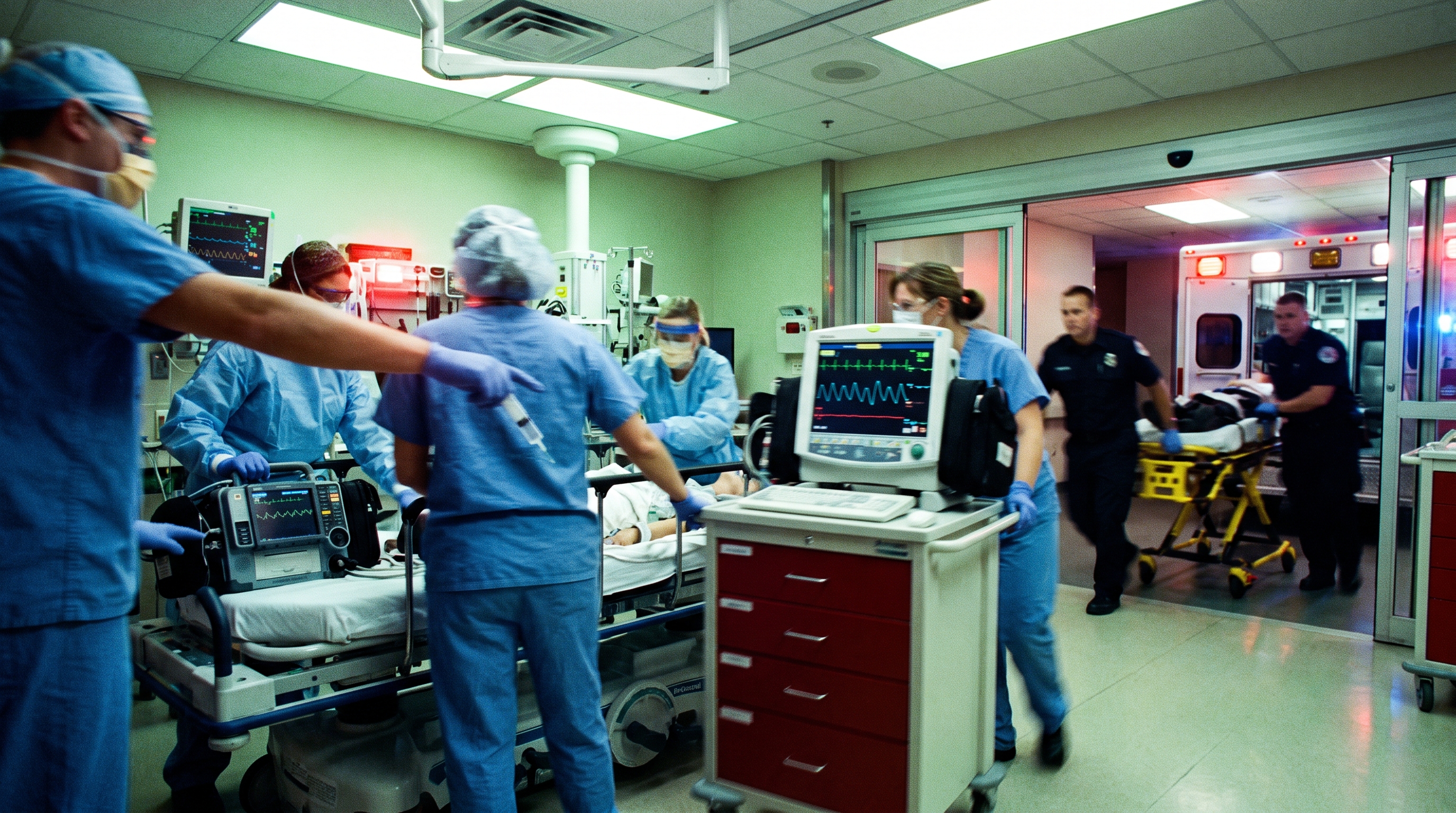
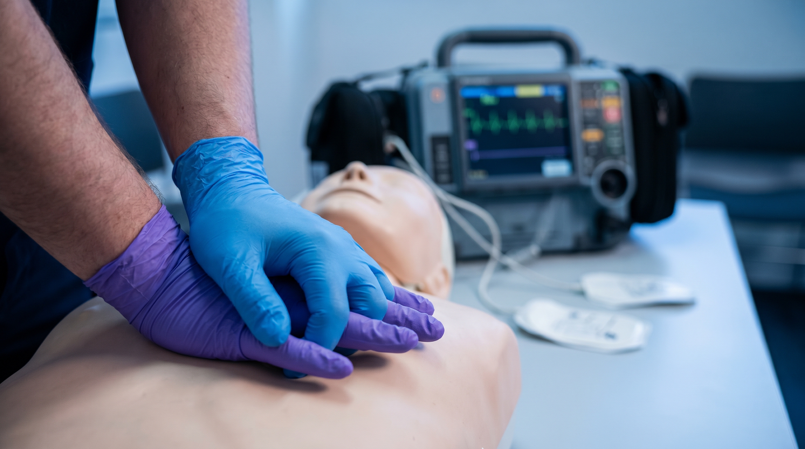
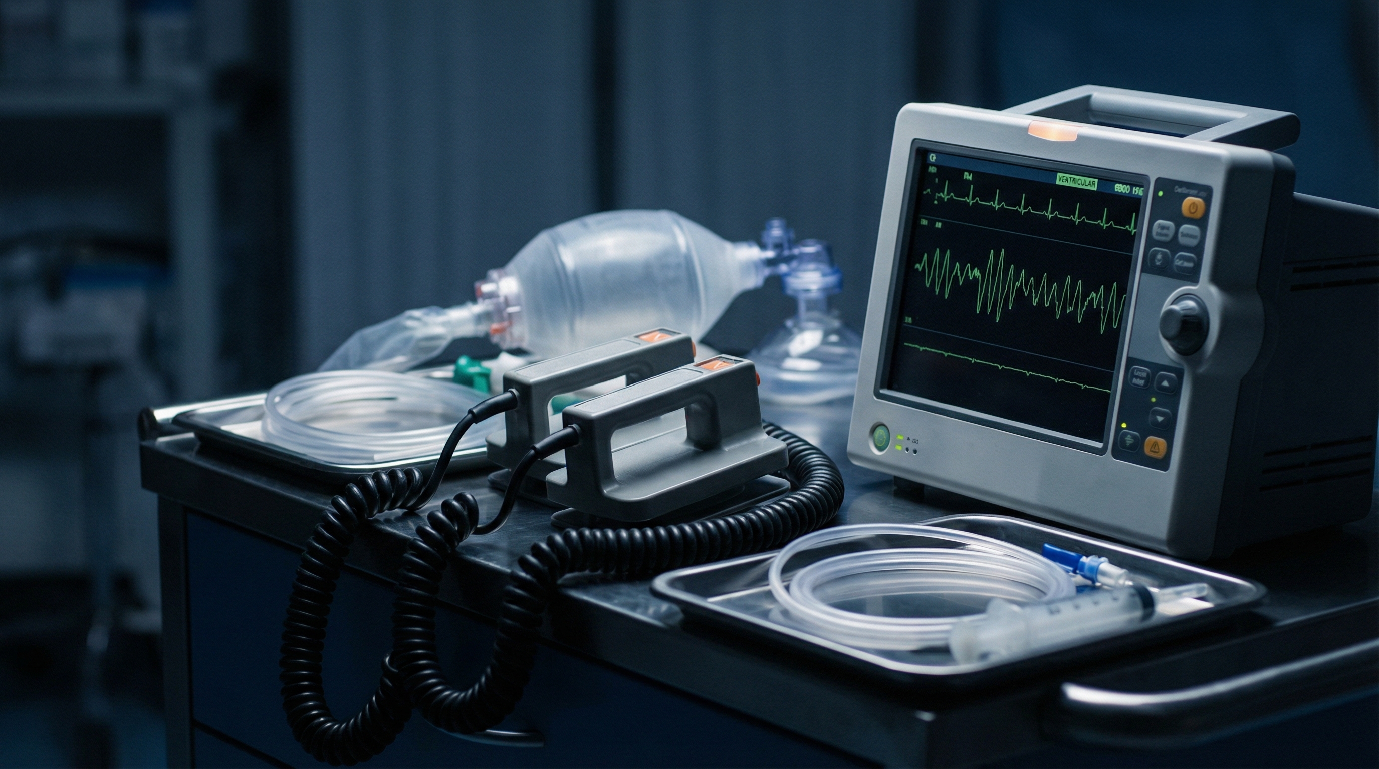
A monitor alarms, the rate reads 180, and the QRS complexes are narrow and regular. You have seconds to decide what to do next, not minutes to page through a manual. That is exactly the moment this decision card is built for: a clean, bedside escalation ladder for stable supraventricular tachycardia (SVT), from the first vagal maneuver through adenosine to synchronized cardioversion.
This card zooms in on a single branch of a much larger algorithm. If you need the full stable-versus-unstable fork and all four tachycardia branches side by side, our adult tachycardia with a pulse algorithm companion guide covers that ground. Here, we stay narrow: what actually works before adenosine, how to push adenosine correctly, when to skip straight to a shock, and the traps that catch even experienced providers. Consider this the working companion to our earlier guides on treating SVT with adenosine and vagal maneuvers before adenosine, distilled into one ladder you can run at the bedside.
Every dose, energy setting, and success rate in this card is drawn from published trial data or current resuscitation references, cited as you go. Nothing here replaces your institution's protocol or medical control, but it should match what you already learned in your ACLS algorithms training almost exactly.

Before any maneuver or medication, confirm three things: the rhythm is regular, the QRS is narrow, generally under 0.12 seconds, and the patient has a pulse with a blood pressure and mental status that can tolerate a minute or two of attempted conversion. This ladder is built for exactly that presentation. An irregular narrow-complex rhythm is more likely atrial fibrillation, atrial flutter with variable conduction, or multifocal atrial tachycardia, and a wide-complex tachycardia, whether regular or irregular, changes the entire approach. We cover why that distinction is so important in the cautions section below, because it is the single most dangerous point on this whole card.
If the patient is unstable, meaning hypotensive, acutely altered, in shock, having active ischemic chest pain, or in acute heart failure because of the rate, do not work through vagal maneuvers and adenosine first. Move directly to Step 4, synchronized cardioversion. Everything between here and there assumes a stable patient.
Say the plan out loud as soon as you commit to it. Announcing "regular, narrow, stable, going to attempt modified Valsalva" gives the rest of the team a chance to flag something you missed, gets a second set of eyes on the monitor, and puts everyone on the same page before a syringe is even drawn up. That habit of closed-loop communication is one of the team dynamics skills every ACLS course drills for a reason: the decision card only works if the whole team is reading it together.
Standard vagal maneuvers, bearing down forcefully while sitting upright, have historically converted SVT to sinus rhythm in only a small fraction of patients. The modified Valsalva technique performs meaningfully better and costs nothing to attempt first. In the multicenter REVERT trial, a modified approach converted 43 percent of patients to sinus rhythm on an intention-to-treat basis, compared with 17 percent for the standard technique. That is roughly a two-and-a-half-fold improvement using nothing but positioning and timing.
The modification is simple enough to teach a patient in real time. Have the patient sit semi-recumbent at roughly 45 degrees and strain into a 10 mL syringe for 15 seconds, generating genuine intra-abdominal pressure rather than a token grunt. Immediately at the end of the strain, lay the patient flat and passively raise both legs to 45 degrees for another 15 seconds before returning them to a seated position. The leg raise augments venous return right as the vagal tone from the strain phase peaks, which is the mechanical reason this version outperforms the classic strain-and-release. Our vagal maneuvers guide walks through the full technique with troubleshooting for patients who cannot generate an effective strain.
Give it one honest attempt, coached well, before moving on. A half-hearted strain is not a failed vagal maneuver, it is a maneuver that was never really tried. If the rhythm does not break and the patient remains stable, move to Step 2.
Most SVT is a reentry circuit that uses the AV node as one leg of a small electrical loop, either entirely within the AV node itself or through an accessory pathway working alongside it. Adenosine binds A1 receptors in the AV node and briefly hyperpolarizes the tissue, producing a transient, near-complete block of conduction through the node. For a fraction of a second, the reentry loop simply has nowhere to go, and if the AV node is genuinely part of the circuit, the tachycardia stops. That mechanism is also why adenosine is diagnostic as well as therapeutic: a rhythm that briefly slows or unmasks flutter waves during the AV block, without ever converting, is telling you the AV node was a bystander, not the circuit itself, per the mechanistic review in StatPearls' overview of adenosine pharmacology.
The same near-instant onset that makes adenosine effective also makes it unforgiving of technique. Its plasma half-life is measured in seconds because red blood cells and vascular endothelium metabolize it almost immediately, which is the entire reason the rapid push-and-flush method exists: a slow push simply never reaches the AV node at an effective concentration.
Adenosine remains the first-line pharmacologic agent for stable, regular, narrow-complex SVT that has not converted with vagal maneuvers. The standard starting adult dose is 6 mg given as a rapid intravenous push, ideally through the largest, most proximal vein available, such as the antecubital fossa, since adenosine's ultra-short half-life means it needs to reach the heart before it is cleared. That 6 mg starting dose, along with the reduced dosing used for central lines, transplant recipients, and interacting medications covered below, is detailed in the dosing summary within StatPearls' adenosine reference.
Technique matters as much as the dose. Draw the adenosine into one syringe and a 20 mL normal saline flush into a second syringe, then attach both to the IV port closest to the patient using a stopcock or Y-connector. Push the adenosine as fast as it will go, over one to three seconds, and immediately follow with the saline flush pushed just as fast, without a pause between the two. Some teams prefer elevating the extremity immediately after the flush to help the bolus reach central circulation faster. If your service uses a single-syringe technique that mixes adenosine and flush together, that approach has also shown comparable success rates in trial data, so use whichever double- or single-syringe method your protocol trains for, as long as the push itself is fast and immediately followed by flush.
Warn the patient before you push, in one sentence: "You are going to feel your heart race, your chest may feel tight or heavy for a few seconds, and you may feel flushed or briefly like something is wrong. This is expected, it passes in under a minute, and it means the medication is working." Patients who are warned tolerate the sensation of transient AV block far better than patients who are not, and they are less likely to panic or pull back from a limb during the push. Keep the monitor running continuously through this, because a brief period of sinus pause or high-grade AV block immediately after the dose is expected, not a complication.
If the rhythm has not converted within one to two minutes of the first dose, give a second dose of 12 mg using the identical rapid push-and-flush technique. Many protocols allow a third dose of 12 mg if the second also fails, though at that point it is worth pausing to reconsider the diagnosis: a narrow-complex tachycardia that will not break with two or three appropriately administered doses of adenosine is more likely to be atrial flutter with fixed conduction, or an atrial tachycardia that is not reentrant and therefore not adenosine-responsive, rather than typical AV nodal reentrant tachycardia.
Keep your dosing reference close at hand during a live event rather than trying to recall exact numbers under pressure. Our ACLS medications cheat sheet lists adenosine alongside every other code-cart drug with dose, route, and indication in one scannable table, which is exactly the kind of reference worth keeping loaded on a phone or clipped to the crash cart.
Two doses that fail to convert a truly regular, narrow-complex, hemodynamically stable SVT is also a reasonable point to loop in cardiology, particularly if the patient has a history of accessory pathway conduction or prior ablation, before pushing a third dose or moving to rate control agents like a calcium channel blocker or beta blocker as an alternative to cardioversion.
Some patients do not get two attempts at adenosine, and some never get one. Move directly to synchronized cardioversion whenever the tachycardia is causing instability: a systolic blood pressure that will not support end-organ perfusion, acute altered mental status, signs of shock, ongoing ischemic chest pain, or acute pulmonary edema that the provider attributes directly to the rate. Adenosine and vagal maneuvers are appropriate for stable patients who can tolerate the minute or two those interventions take; an unstable patient cannot.
For a regular, narrow-complex rhythm like SVT, current teaching favors starting at a relatively low synchronized energy, commonly in the 50 to 100 joule range on a biphasic defibrillator, and doubling the energy on subsequent attempts if the first shock fails to convert the rhythm, as summarized in StatPearls' review of synchronized electrical cardioversion. Always confirm the defibrillator is in synchronized mode before delivering the shock, since an unsynchronized shock landing on a T wave can precipitate ventricular fibrillation instead of correcting the rhythm in front of you. Our synchronized cardioversion guide walks through pad placement, mode confirmation, and troubleshooting a machine that will not sync.
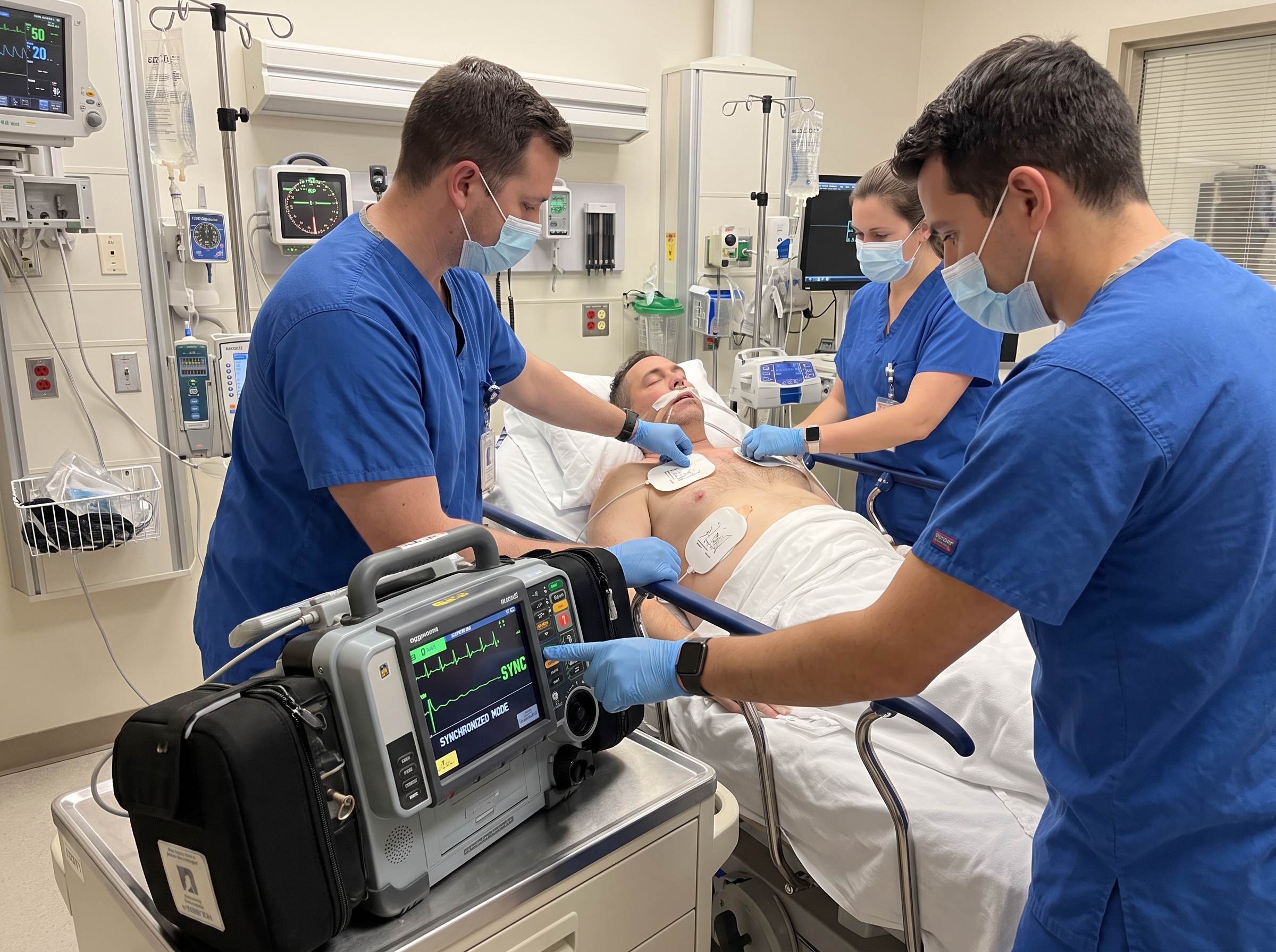
If the patient has any tolerable window at all, even thirty seconds, use it for procedural sedation rather than shocking a conscious patient without warning. Our conscious sedation for cardioversion guide covers agent selection and monitoring for exactly this scenario. When there truly is no window, because the patient is critically unstable, cardioversion proceeds without waiting for sedation to take effect.
A handful of clinical situations change this ladder meaningfully. Four are worth committing to memory before you are standing at the bedside.
This is exactly why Step 0 insists on confirming a regular, narrow rhythm before you climb any further up this ladder. When the rhythm will not commit to being clearly regular and narrow, slow down, get a 12-lead if the patient's stability allows it, and treat any doubt about pre-excitation as a reason to avoid adenosine rather than a reason to try it and see.
Once the rhythm converts, whether from a vagal maneuver, adenosine, or a shock, get a 12-lead ECG in sinus rhythm as soon as it is practical. This is the best chance to spot a delta wave or short PR interval suggesting an underlying accessory pathway, a finding that changes both the patient's long-term management and how you would approach the next episode. Keep the patient on continuous monitoring for a reasonable observation period, since a brief run of recurrent SVT immediately after conversion is not unusual, particularly after adenosine, which clears the system within roughly ten seconds and offers no lingering protection against reinitiation.
Recheck a full set of vital signs once the rhythm is confirmed sinus, rather than assuming stability just because the monitor rate looks normal again. Some patients run transiently hypotensive right after conversion, particularly following cardioversion under sedation, and that window deserves its own attention before you move on to disposition planning.
Document the rhythm strips from before, during, and after each intervention, including the exact vagal maneuver technique used, each medication dose and time, and the sync mode and energy for any shock delivered. That record matters for the cardiology follow-up and for anyone reviewing the case afterward.
Patients with recurrent SVT, particularly those who have needed adenosine or cardioversion more than once, are reasonable candidates for outpatient cardiology or electrophysiology referral rather than repeat emergency visits. Catheter ablation targeting the reentrant circuit, whether AV nodal reentry or an accessory pathway, has a well-established track record for preventing recurrence in appropriately selected patients, and it is worth raising as an option rather than treating every episode as an isolated event to be broken and discharged.
In the meantime, some patients benefit from being taught the modified Valsalva technique themselves, so they have a first-line, zero-risk option to try at home before an episode escalates to the point of needing emergency care. That single piece of patient education, handed over at discharge, is one of the more useful five-minute investments you can make in a patient who has just gone through this ladder once and does not want to repeat the experience.
It is also worth asking about common triggers before discharge: caffeine intake, alcohol, sleep deprivation, dehydration, and stimulant use all lower the threshold for reentrant tachycardia in susceptible patients. None of that replaces a cardiology referral for someone with true recurrent SVT, but it gives the patient something concrete to modify while they wait for that appointment.
A decision card like this one is meant to be reviewed cold, not read for the first time while a monitor is alarming. If it has been a while since you last worked through the full tachycardia algorithm, adenosine dosing, and synchronized cardioversion technique together under exam conditions, that gap is worth closing before it closes itself at the worst possible moment.
An ACLS certification or recertification course built by practicing emergency physicians walks through this exact ladder, along with the rest of the tachycardia and bradycardia algorithms, at your own pace and on your own schedule. Courses are self-paced with no time limit, include unlimited retakes on the exam at no extra charge, and come with a money-back guarantee if your employer will not accept the certification, so there is little downside to using the course as your refresher rather than relying on memory alone.
.jpg)