A patient's monitor throws up an irregular rhythm, the PR interval looks "off," and someone at the bedside asks the question that matters most: is this dangerous, and what do I do about it right now? Heart blocks have a reputation for being one of the trickiest calls in rhythm recognition, but the classification itself is not complicated. What makes it feel hard is trying to reason through pathophysiology while a patient is symptomatic in front of you.
This guide is built for that moment. It is the fast, scannable version of AV block recognition, organized the way you actually need it during a code or a bedside decompensation: how to sort first, second, and third degree block in about ten seconds, which ones are benign versus dangerous, and exactly where each one lands in the ACLS bradycardia algorithm. If you want the deeper pathophysiology, conduction system anatomy, and etiology behind each block, our comprehensive guide to atrioventricular blocks covers that in detail. This article assumes you already know the difference between a P wave and a QRS complex; if rhythm basics feel shaky, start with our beginner's guide to core ACLS heart rhythms first.

Before you memorize definitions, memorize the process. Every AV block can be sorted with three quick questions, answered by looking at a run of consecutive beats rather than a single complex.
Walking that decision tree takes less time to do than to read. The key habit is looking at a strip of six to ten beats, not just one complex, because the pattern only reveals itself across multiple cycles. This same pattern-recognition skill is exactly what ACLS certification is designed to build until it is automatic under pressure; our ACLS certification course walks through rhythm recognition and the bradycardia algorithm together so the two skills reinforce each other.
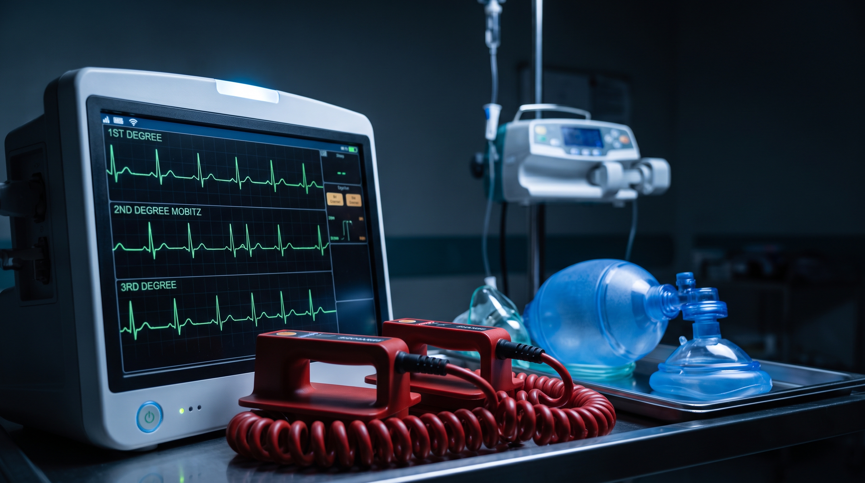
First degree AV block is the simplest of the four patterns. Every P wave is followed by a QRS complex, one to one, with no dropped beats at all. The only abnormality is timing: the PR interval is prolonged beyond the normal upper limit, and according to StatPearls, that threshold is a PR interval greater than 0.20 seconds, or one large box on standard ECG paper.
Clinically, first degree block is almost always a bystander finding rather than the reason a patient is unstable. It reflects a conduction delay somewhere between the atria and the ventricles, most often within the AV node itself, and it is common in healthy people with high vagal tone, athletes, and patients on AV-nodal-blocking medications like beta blockers or calcium channel blockers. On its own, first degree block rarely requires treatment. The exception worth remembering for the algorithm is a markedly prolonged PR interval, sometimes called "first degree block plus," which can cause loss of AV synchrony severe enough to produce fatigue or exercise intolerance even though the rhythm technically still conducts every beat.
Second degree Mobitz I, better known as Wenckebach, is the pattern most providers find satisfying once they see it: the PR interval lengthens progressively, beat after beat, until one P wave simply fails to conduct and the cycle resets. Because the PR intervals grow before the drop, the R-R interval actually shortens slightly with each conducted beat, then pauses at the dropped beat. This creates the classic "grouped beating" appearance on a strip, where clusters of QRS complexes are separated by a gap.
Mobitz I almost always arises from a conduction delay within the AV node itself, which is a more forgiving location than what you will see in Mobitz II. It shows up in the same populations as first degree block: high vagal tone, inferior wall ischemia, digoxin effect, and AV-nodal-blocking drugs. Most patients with Wenckebach are asymptomatic and hemodynamically stable, and the rhythm often resolves on its own once the underlying cause, such as a medication or transient vagal surge, is addressed. It only becomes an acute concern when the ventricular rate drops low enough to cause symptoms, which shifts management toward the same instability criteria used for any symptomatic bradycardia.
Mobitz II looks deceptively similar to Mobitz I at first glance, since both involve intermittently dropped QRS complexes. The difference is that in Mobitz II, the PR interval stays fixed and identical on every single conducted beat, right up until a P wave suddenly fails to conduct with no warning at all. There is no progressive lengthening to tip you off, which is exactly why it is dangerous: the next dropped beat, or the next several in a row, cannot be predicted from the strip in front of you.
The reason Mobitz II carries so much more risk comes down to location. Where Mobitz I typically originates in the AV node, Mobitz II usually reflects disease in the His-Purkinje system, below the AV node, often accompanying a widened QRS from an underlying bundle branch block. A review in PMC notes that Mobitz II is both commonly overdiagnosed and clinically important to identify correctly, since true Mobitz II carries a meaningfully higher risk of sudden, unpredictable progression to complete heart block than Mobitz I does. StatPearls reinforces that Mobitz II is considered pathologic essentially by definition and warrants a more aggressive posture even in a patient who currently looks stable, because the underlying conduction system is structurally diseased rather than transiently slowed.
If you only remember one thing under pressure, remember this: measure the PR interval on three or four conducted beats in a row before the dropped beat. If the PR interval is changing, growing longer with each beat, you are looking at Mobitz I. If the PR interval is identical, beat after beat, right up until the sudden drop, you are looking at Mobitz II. A wide QRS complex on the conducted beats is another clue that tips the suspicion toward Mobitz II and an infranodal problem, since a healthy His-Purkinje system typically conducts with a narrow QRS.
Third degree, or complete, heart block is the most severe pattern on the spectrum. There is a total breakdown in communication between the atria and the ventricles: P waves march along at their own regular rate, QRS complexes march along at their own separate rate, and the two have no consistent relationship to each other whatsoever. This is true AV dissociation. Per StatPearls, since the atrial impulse never reaches the ventricles, cardiac output depends entirely on whatever escape rhythm the ventricles can generate on their own.
That escape rhythm is the clue worth training your eye to catch, because it tells you how much reserve the patient has left. A junctional escape rhythm, narrow QRS, rate roughly 40 to 60 beats per minute, is more stable and comes from a backup pacemaker closer to the AV node. A ventricular escape rhythm, wide QRS, rate often in the 20s or 30s, is far less reliable and can fail outright with no further warning. Complete heart block can be fatal without prompt recognition and treatment, which makes it the pattern where hesitation costs the most.
It helps to think of the four patterns on a spectrum rather than four unrelated diagnoses. First degree block and Mobitz I are usually AV nodal, usually benign, and usually reversible once the underlying trigger is addressed. Mobitz II and third degree block are usually infranodal, usually structural, and carry real risk of sudden hemodynamic collapse even in a patient who looks fine on first assessment.
That said, the rhythm classification alone does not decide the treatment path. What decides it is whether the patient is stable or unstable, exactly as outlined in the ACLS bradycardia algorithm. A patient in Mobitz I with a rate of 58 and no symptoms needs monitoring, not intervention. A patient in Mobitz II with hypotension, altered mental status, signs of shock, ischemic chest discomfort, or acute heart failure needs immediate action regardless of how "textbook mild" the rhythm classification sounds. Signs of instability are what move a patient from observation into active treatment, and a high-grade block should raise your index of suspicion even before instability is obvious, since these rhythms can decompensate quickly.
Once you know the classification and the stability picture, the algorithm path follows logically.
Our ACLS Algorithms hub lays out the complete bradycardia decision tree alongside every other core algorithm, so you can cross-reference the exact drug doses and decision points in one place while you study.
This is the piece that trips people up on exams and at the bedside alike. Atropine works by blocking vagal tone at the SA node and the AV node, which speeds conduction through those structures. That mechanism is a great fit for a nodal problem like Mobitz I. It is a poor fit for Mobitz II or third degree block with a wide escape QRS, because the diseased tissue causing the block sits below the AV node, in the His-Purkinje system, where vagal tone has little influence. Giving atropine to a patient with true infranodal block can do nothing, or in some reported cases can paradoxically increase the atrial rate while the ventricular rate stays just as slow, worsening the ratio of blocked beats. This is exactly why the algorithm favors moving straight to transcutaneous pacing for high-grade block instead of cycling through repeated doses of a drug that was never going to reach the site of the problem. If you want a deeper walkthrough of pad placement, capture thresholds, and troubleshooting a pacer that is not capturing, see our guide to transcutaneous pacing.

Save this table as your quick-reference card. It condenses everything above into the five facts you actually need mid-shift.
Block Type PR Interval Pattern P-to-QRS Relationship Danger Level ACLS Action First Degree Fixed, prolonged beyond 0.20 sec Every P conducts, 1:1 Low; usually a bystander finding Monitor; treat any reversible cause Second Degree, Mobitz I (Wenckebach) Progressively lengthens, then a beat drops Predictable grouped beating Usually low; nodal and often reversible Observe if stable; atropine if symptomatic Second Degree, Mobitz II Fixed on conducted beats, sudden unpredictable drop No warning before a dropped QRS High; infranodal, can progress suddenly Pacer pads on; transcutaneous pacing first line if symptomatic Third Degree (Complete) No fixed relationship; PR is meaningless here Full AV dissociation; independent rates Highest; escape rhythm may fail Pacing first line; atropine unlikely to help
Heart blocks reward exactly the kind of repeated, deliberate strip practice that turns a hesitant guess into an instant call. The three-question method in this guide gets you to a classification fast; understanding why Mobitz II and third degree block resist atropine is what keeps you from wasting critical minutes on a drug that was never going to work. For the pathophysiology behind why each block occurs where it does, revisit our comprehensive AV blocks guide, and pair this article with our printable ACLS rhythm recognition cheat sheet to keep every core rhythm, not just AV blocks, at your fingertips.
If your certification or recertification is on the calendar, rhythm recognition like this is exactly what an ACLS course should drill until it feels automatic rather than academic. Affordable ACLS was built by practicing emergency physicians specifically so that studying decision-making, not memorizing trivia, is the whole point.
.jpg)