When a patient goes into cardiac arrest, every second of resuscitation counts — but not all causes of arrest are created equal. Among the ten reversible causes embedded in the ACLS framework's H's and T's, hypoxia stands in a category of its own. Unlike tension pneumothorax or cardiac tamponade, which require procedural interventions, or toxicological arrests that demand specific antidotes, hypoxia can often be corrected with nothing more than a well-placed mask and effective ventilation. That simplicity is deceptive, though: hypoxia is simultaneously the most common reversible cause of cardiac arrest and one of the most frequently mismanaged during resuscitation.
For any healthcare provider working a code, the phrase "airway-first mindset" is more than a mnemonic device — it's a clinical imperative. In this article, we break down the physiology of hypoxic cardiac arrest, walk through the evidence around airway management during resuscitation, and provide practical guidance for building the reflexes that let you address the airway without sacrificing CPR quality or losing time to the wrong intervention. Whether you're a new nurse stepping into your first code or an experienced clinician brushing up before recertification, understanding hypoxia's central role in reversible cardiac arrest is non-negotiable.
Hypoxia, by definition, is inadequate oxygen delivery to tissues. In the context of cardiac arrest, what we're most concerned with is hypoxemia — dangerously low arterial oxygen content — and its downstream effect on myocardial function. The timeline from respiratory arrest to cardiac arrest is brutally short. When breathing stops or becomes severely compromised, oxygen saturation begins to fall within seconds. Without intervention, the heart typically arrests within three to five minutes.
The mechanism unfolds in a predictable sequence. As PaO2 drops, the myocardium becomes increasingly vulnerable. Hypoxia causes cellular ATP depletion, which disrupts ion channel function and destabilizes membrane potentials. The result is a milieu perfectly suited for malignant dysrhythmias — initially bradycardia and ectopic beats, then ventricular fibrillation or asystole. In pediatric patients, this sequence is even more pronounced: children are far more likely than adults to arrive at cardiac arrest through primary respiratory failure, making hypoxia correction the cornerstone of pediatric resuscitation curricula like PALS.
Importantly, the relationship runs in both directions. Hypoxia causes cardiac arrest, but cardiac arrest perpetuates hypoxia. Once circulation ceases, passive oxygen delivery stops entirely. Every minute of untreated cardiac arrest compounds the hypoxic insult to the brain and other vital organs. This bidirectional relationship is why the ACLS algorithm demands both high-quality chest compressions and early ventilation — neither alone is sufficient to break the cycle.
The H's and T's represent the ACLS provider's systematic approach to reversible causes of cardiac arrest. This framework is most critical during pulseless electrical activity (PEA) and asystole, where organized electrical activity exists but no effective perfusion occurs. When you see PEA on the monitor, your diagnostic mindset must immediately shift toward identifying which underlying condition is preventing the heart from translating electrical activity into mechanical output.
The five H's are hypovolemia, hypoxia, hydrogen ion excess (acidosis), hypo- or hyperkalemia, and hypothermia. The four T's are tension pneumothorax, tamponade (cardiac), toxins, and thrombosis (pulmonary or coronary). Of these, hypoxia occupies a unique position: it is at once a direct cause of arrest, a complication of ongoing resuscitation, and a consequence of virtually every other reversible cause. A patient in tension pneumothorax is also hypoxic. A severely hypovolemic patient will become hypoxic. A patient in opioid toxicity stops breathing and becomes profoundly hypoxic before the heart stops. In this sense, addressing the airway and correcting hypoxia is a near-universal intervention regardless of the underlying etiology.
For a comprehensive breakdown of all ten reversible causes and how to systematically work through them at the bedside, see our guide on Sudden Cardiac Arrest: The Hs and Ts You Need to Know. Understanding where hypoxia fits within the full differential is the first step toward a structured, efficient code response.
One of the most clinically valuable skills any ACLS provider can develop is the ability to recognize impending respiratory arrest before it completes the journey to cardiac arrest. The window between respiratory decompensation and cardiac standstill is brief, but it is real — and acting within it dramatically changes patient outcomes. According to the Merck Manual, cardiac arrest follows uncorrected respiratory arrest within minutes, driven by the combined effects of hypoxemia and hypercapnia.
The clinical signs of impending respiratory arrest are not subtle once you know what to look for: altered mental status, cyanosis, decreasing respiratory rate, agonal breathing, use of accessory muscles, poor air movement on auscultation, and declining oxygen saturation despite supplemental oxygen. Any of these signs should trigger immediate escalation of airway management — not observation, not a wait-and-see approach, but direct intervention.
For healthcare providers who want to deepen their understanding of this critical clinical state and how to intervene effectively before the patient codes, our detailed reference on Managing Respiratory Arrest: A Comprehensive Guide covers assessment, intervention priorities, and escalation pathways in granular detail.
For decades, ACLS training emphasized early endotracheal intubation as the gold standard for airway management in cardiac arrest. The reasoning made physiological sense: a secured, definitive airway should allow better oxygenation and ventilation than a mask held against a face. The clinical evidence, however, has complicated this narrative considerably.
A landmark randomized clinical trial published in JAMA compared bag-mask ventilation to endotracheal intubation during out-of-hospital cardiac arrest and found no significant difference in neurological outcomes at 28 days — 4.3% favorable outcomes in the bag-mask group versus 4.2% in the intubation group. A subsequent large multicenter study published in Circulation further supported bag-valve-mask ventilation as a viable strategy associated with comparable survival outcomes.
These findings carry an important practical implication: attempting intubation introduces hands-off time, interrupts chest compressions, and can delay the delivery of epinephrine. If the intubation is difficult or unsuccessful, it can transform a manageable situation into a catastrophic one. The 2025 AHA Guidelines reflect this shift in thinking, reinforcing that high-quality bag-mask ventilation, applied correctly and consistently, is a legitimate and evidence-supported primary airway strategy in many cardiac arrest scenarios.
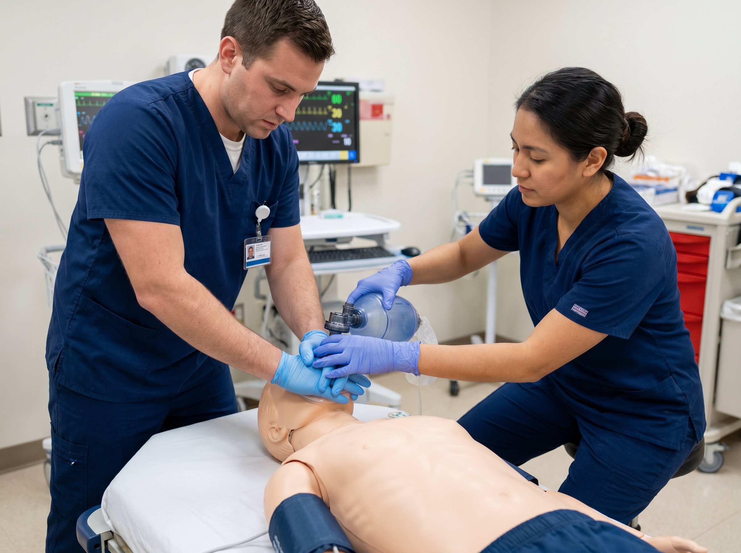
Correct technique is essential. Two-person bag-mask ventilation — with one provider forming a seal using both hands and another squeezing the bag — consistently outperforms single-person technique in seal quality and delivered tidal volume. Providers should aim for a tidal volume that produces visible chest rise, typically 500 to 600 mL in adults, and should ventilate at 10 breaths per minute once an advanced airway is in place, or in a 30:2 ratio during CPR without an advanced airway. For a thorough review of the mechanics and common pitfalls of this foundational skill, see our guide on Mastering Bag and Mask Ventilation.
Bag-mask ventilation is a highly effective first-line intervention, but it is not always sufficient. Certain patient populations and clinical scenarios warrant earlier escalation to advanced airway management. These include patients with massive emesis, anatomical barriers to effective mask seal, severe burns or trauma to the face and upper airway, prolonged resuscitation where sustained ventilation quality is declining, or situations where a single provider cannot maintain an adequate seal while also supporting compressions.
When advanced airway placement is indicated, modern ACLS practice offers two primary options: supraglottic airways (SADs) such as the i-gel or laryngeal mask airway (LMA) and endotracheal intubation (ETI). Neither approach is categorically superior in the out-of-hospital setting, and the 2025 guidelines avoid declaring a universal preference. The right choice depends on operator experience, available equipment, patient anatomy, and clinical context.
Supraglottic airways have become increasingly favored in out-of-hospital and resource-limited environments because they can be placed quickly, require less training to achieve competency, and allow uninterrupted chest compressions during insertion. Endotracheal intubation remains the preferred advanced airway when a skilled operator is available and when definitive airway protection — such as in aspiration risk or expected prolonged post-arrest care — is a priority. For a detailed clinical comparison of these two approaches, including placement technique and confirmation methods, refer to our resource on Supraglottic Airways in ACLS: When to Choose i-gel or LMA Over Endotracheal Intubation.
No discussion of airway management in cardiac arrest is complete without addressing capnography. End-tidal CO2 (ETCO2) monitoring via waveform capnography has become one of the most powerful tools in the modern resuscitator's arsenal — not just for confirming airway placement, but for tracking the effectiveness of CPR and anticipating return of spontaneous circulation (ROSC).
When an advanced airway is in place, ETCO2 monitoring provides continuous feedback on ventilation effectiveness and cardiac output. During CPR, ETCO2 values correlate with cardiac output generated by chest compressions: higher ETCO2 means better compressions and better forward flow. A sudden, sustained rise in ETCO2 — often jumping from baseline values of 10 to 20 mmHg to 35 to 40 mmHg — is one of the most reliable early indicators of ROSC, often preceding a palpable pulse by seconds. Conversely, persistently low ETCO2 values below 10 mmHg despite optimal CPR technique are associated with poor prognosis and may inform shared decision-making about resuscitation duration.

Waveform capnography also instantly detects esophageal intubation — a potentially fatal complication of ETI — by demonstrating a flat waveform with CO2 readings near zero. No other confirmation method is as reliable. The 2025 AHA guidelines strongly recommend ETCO2 monitoring as a quality assurance tool throughout resuscitation. For a deep-dive into how to interpret waveforms and apply ETCO2 data in real-time clinical decision-making, see Capnography in Cardiac Arrest: How End-Tidal CO2 Monitoring Guides ACLS Decision-Making.
Pulseless electrical activity (PEA) is one of the most diagnostically challenging rhythms in ACLS because it presents with organized electrical activity but no effective pulse. The temptation is to follow a purely pharmacological approach — epinephrine every 3 to 5 minutes, CPR, rhythm check — but without identifying and treating the underlying cause, even perfect ACLS execution may fail to achieve ROSC.
Hypoxic PEA presents a specific pattern worth recognizing: it most commonly arises from a respiratory precursor event, such as respiratory failure, aspiration, or severe asthma exacerbation. In these patients, effective airway management and oxygenation can produce dramatic and rapid improvement. Unlike hemorrhagic or obstructive causes of PEA, where resuscitation may require blood products, needle decompression, or pericardiocentesis, hypoxic PEA responds to ventilation — sometimes within a single cycle of high-quality CPR accompanied by effective bag-mask ventilation.
Clinical clues that hypoxia may be driving a PEA arrest include: a witnessed respiratory event preceding arrest, a clinical history of airway compromise such as anaphylaxis, drowning, opioid overdose, or severe asthma, prolonged unresponsiveness before EMS arrival, or cyanosis on exam. In these scenarios, aggressive airway management is not a secondary consideration — it is the primary intervention. For a comprehensive review of PEA recognition and the full spectrum of reversible causes, explore our resource on Understanding Pulseless Electrical Activity (PEA): Causes and Treatment.
Clinical knowledge is only as good as the reflexes it produces under pressure. The airway-first mindset is not a suggestion to delay CPR — chest compressions remain the foundation of every cardiac arrest response. Rather, it is a systematic cognitive priority that ensures airway management is never an afterthought, never delegated to the least experienced member of the team, and never delayed in favor of procedures that can wait.
Here is a practical framework for integrating an airway-first mindset into your code response:
The 2025 AHA Guidelines for CPR and Emergency Cardiovascular Care represent the most current evidence synthesis available to practicing clinicians. Regarding airway management, the 2025 AHA Advanced Life Support guidelines reinforce that airway management decisions should be individualized based on provider skill, available equipment, and patient factors — with no single advanced airway technique recommended universally over others.
On the research frontier, a 2025 study examining the apnea interval — the time during which ventilation is interrupted for tracheal intubation — found that prolonged apnea intervals are associated with worse outcomes in cardiac arrest resuscitation, reinforcing the argument for minimizing airway management time and prioritizing techniques that can be performed rapidly without prolonged CPR interruption. This research, published in the journal Resuscitation, underscores why provider skill with both BVM and supraglottic airways is essential — the best advanced airway is the one that can be placed quickly and confidently.
The StatPearls comprehensive review of ACLS further reinforces that among the H's and T's, hypoxia demands immediate procedural response — airway management is not a supportive measure but a primary therapeutic intervention. Providers who treat ventilation as secondary to pharmacology in hypoxic arrest are misallocating their resuscitation resources.
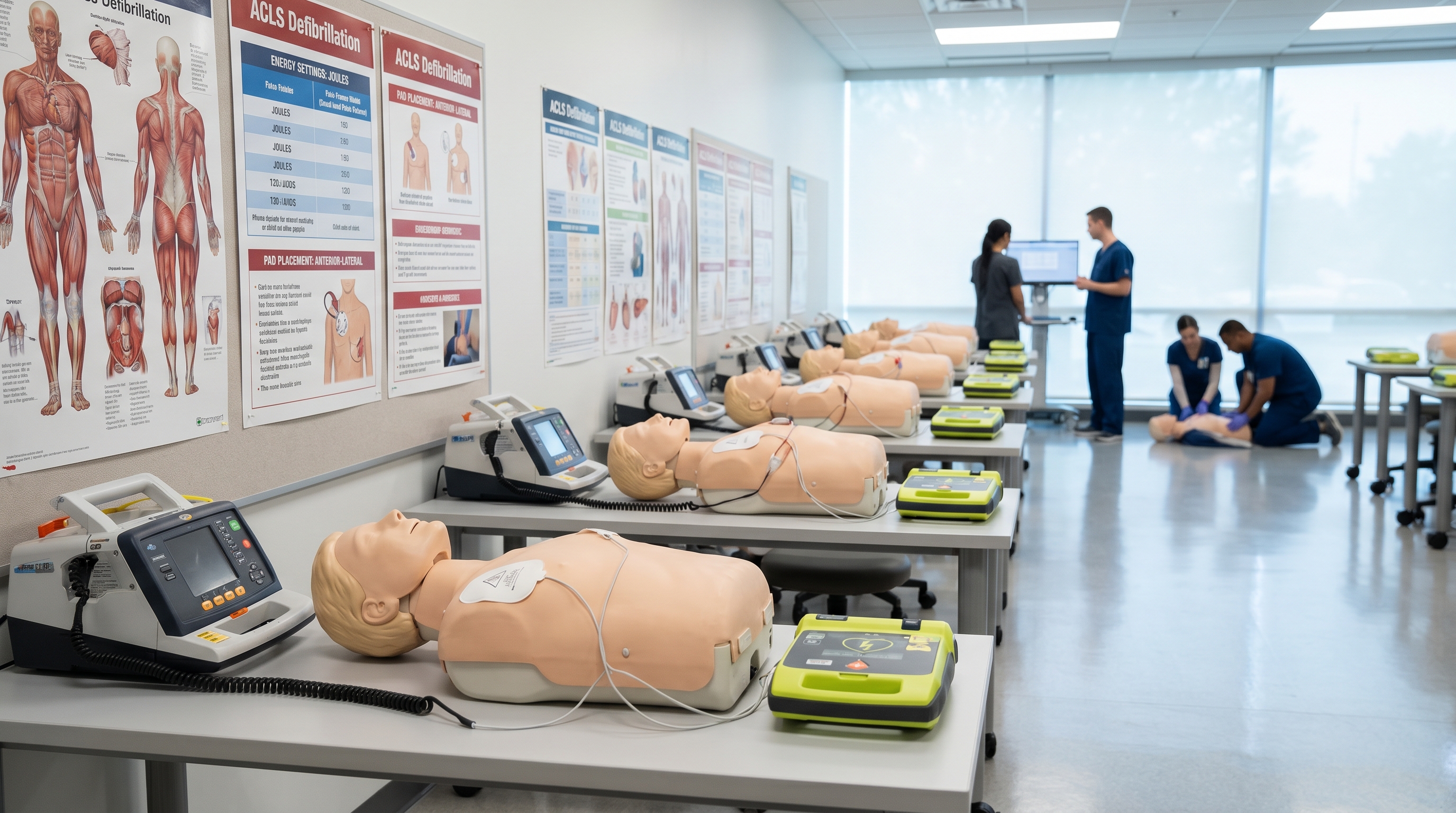
Knowing the physiology and protocols is necessary but not sufficient. The airway-first mindset must be rehearsed until it becomes automatic under the physiological stress of a real code. This is precisely where structured ACLS certification provides value that no amount of passive reading can replicate.
ACLS training builds cognitive rehearsal for the systematic assessment of reversible causes, including hypoxia. It reinforces the role assignments that prevent airway management from being overlooked during a chaotic resuscitation. It develops the technical confidence to perform two-person BVM effectively, recognize ETCO2 waveforms, and make the intubation-versus-supraglottic-airway decision under time pressure. For healthcare providers who need to certify or recertify, the platform should offer current, evidence-based content that reflects the 2025 AHA guidelines — not outdated curricula.
At Affordable ACLS, our online ACLS certification course is developed by board-certified emergency medicine physicians with more than 20 years of combined clinical and academic experience. The course covers the full spectrum of ACLS content — including the H's and T's, airway management strategies, capnography interpretation, and PEA management — at a fraction of the cost of traditional in-person programs. Courses are self-paced, completed in one to two hours, and provide immediate downloadable certification upon passing. ACLS certification is available for $99, with recertification at $89. For providers who also need BLS or PALS credentials, bundle pricing brings the total cost down further.
Every member of your team who might respond to a code should understand hypoxia's role in cardiac arrest — not as a background fact but as an actionable clinical priority. Whether you're building a new team from scratch or ensuring your existing team's certifications reflect the latest evidence, online certification offers the flexibility to train on your schedule without sacrificing clinical rigor. When you're ready to certify, our guide to key ACLS guideline changes for 2025 is an excellent way to ensure your foundational knowledge is current before you sit the exam.
Among all the interventions available during a cardiac arrest — epinephrine, amiodarone, defibrillation, intraosseous access — none addresses the most common reversible cause more directly than securing the airway and delivering oxygen. Hypoxia is simultaneously the most preventable precursor to cardiac arrest and the most correctable driver of PEA and asystole once arrest occurs.
The airway-first mindset does not compete with compressions-first or rhythm-first approaches. It integrates with them. High-quality CPR restores coronary perfusion pressure. Defibrillation terminates shockable rhythms. But neither works optimally in the presence of uncorrected hypoxia. Oxygen delivery — achieved through whatever airway technique is most effective for the individual provider and patient — must be a parallel priority, not a sequential afterthought.
If you are a healthcare professional who responds to codes, the question is not whether hypoxia will present itself in your resuscitation career. It will. The question is whether you will recognize it early, intervene decisively, and apply the airway management skills that give your patient the best possible chance at ROSC and a meaningful neurological recovery. ACLS certification through a physician-developed, evidence-based curriculum is the structured foundation for building exactly that competence. To learn more about certifying with Affordable ACLS and ensuring your airway skills reflect the latest clinical evidence, visit AffordableACLS.com or contact our support team at 866-655-2157.
.jpg)