ACLS Blogs

ACLS Tachycardia Algorithm Companion: Stable vs Unstable Decisions

The Tachycardia Algorithm Is a Decision Tree, Not a Rhythm List

Most providers picture the ACLS tachycardia algorithm as a rhythm strip quiz: identify the arrhythmia, then treat it. That is not how the algorithm is built. The Adult Tachycardia with a Pulse Algorithm is a branching decision tool, and every branch point is a question about the patient in front of you, not a question about the ECG in isolation. Get the branching logic right and the specific rhythm underneath becomes much easier to manage. Get it wrong, and you can shock a patient who did not need it or waste critical minutes on a stable-appearing patient who is actually crashing.


This companion walks through the algorithm the way it is actually built: first the stability call, then the fork between the unstable and stable branches, then the four boxes that stable tachycardia splits into based on QRS width and rhythm regularity. If you want the rhythm-by-rhythm mechanics of narrow-complex SVT specifically, including vagal maneuvers and adenosine dosing, that lives in its own decision card. Here, the goal is to make sure you never lose the thread of the algorithm's logic, even when the rhythm strip in front of you looks unfamiliar.

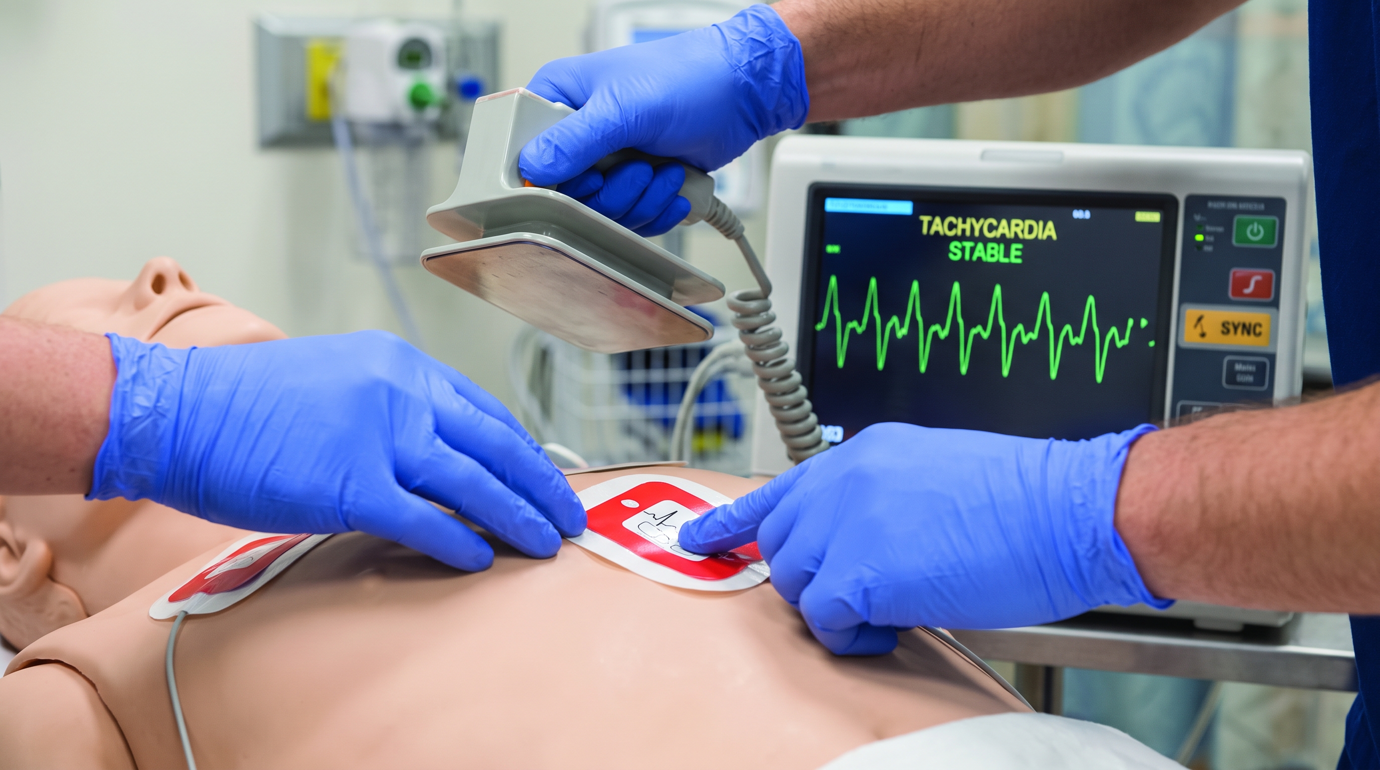
Emergency care team assessing a patient's cardiac monitor to judge hemodynamic stability during tachycardia


Step One: Is the Patient Stable or Unstable

Every pass through the tachycardia algorithm starts the same way: identify and treat the underlying cause, then assess whether the tachyarrhythmia itself is causing hemodynamic compromise. The algorithm defines instability using a specific, limited list of findings. A patient is considered unstable when the tachycardia is producing one or more of the following: hypotension, altered mental status, signs of shock, ischemic chest discomfort, or acute heart failure.


  • Hypotension - a blood pressure that is inadequate to perfuse the brain, kidneys, and myocardium
  • Altered mental status - new confusion, agitation, or decreased responsiveness that was not present before the rhythm change
  • Signs of shock - cool, pale, clammy skin, delayed capillary refill, or a weak thready pulse pointing to poor peripheral perfusion
  • Ischemic chest discomfort - chest pain or pressure consistent with myocardial ischemia that appeared or worsened with the fast rate
  • Acute heart failure - flash pulmonary edema, jugular venous distention, or new respiratory distress tied to the arrhythmia


Notice what is missing from that list: the heart rate itself. A number on the monitor is not, by itself, a criterion for instability. This is the single most common trap in tachycardia management, both in the resuscitation bay and on the certification exam. A young, otherwise healthy patient with a narrow-complex rhythm at 160 beats per minute who is talking to you, mentating normally, and maintaining a normal blood pressure is not unstable just because the number looks alarming. Conversely, a rate of 130 in a frail patient with new hypotension and confusion absolutely qualifies as unstable. The rate sets the stage; the clinical findings make the call.


Is the Tachycardia Causing the Instability, or Is Something Else Driving the Rate

The second trap sits right next to the first one, and it is the piece of reasoning that trips up exam candidates most often: before you treat the rate as the primary problem, ask whether the tachycardia is the cause of the instability or a compensatory response to something else. Sinus tachycardia from sepsis, hemorrhage, pain, fever, hypovolemia, or pulmonary embolism can produce a fast, sometimes irregular-looking rhythm alongside genuine hypotension or altered mental status. That patient is not a synchronized cardioversion candidate. Shocking sinus tachycardia does nothing for the underlying problem and can delay the fluids, blood, or source control the patient actually needs.


The distinction usually comes down to context and rhythm morphology rather than the rate alone. Sinus tachycardia rarely exceeds the physiologic maximum for age, tends to have an identifiable P wave before every QRS complex, and gradually speeds up or slows down rather than switching on abruptly. A primary tachyarrhythmia such as SVT, atrial fibrillation with rapid ventricular response, atrial flutter, or ventricular tachycardia is more likely to have an abrupt onset, a regular and unusually fast rate for a compensatory response, or a QRS morphology that does not fit a sinus mechanism. When the history and exam suggest an obvious driver such as fever, blood loss, or dehydration, treat that driver first and reassess the rhythm before reaching for the algorithm's unstable branch.


The Unstable Branch: Straight to Synchronized Cardioversion

Once you have confirmed that the tachyarrhythmia itself is the cause of the instability, the algorithm does not offer a menu of options. The unstable branch goes directly to synchronized cardioversion. There is no antiarrhythmic infusion to try first, no vagal maneuver to attempt, no waiting to see if the rate comes down on its own. Sedation should be given whenever the patient's condition allows, but the priority is restoring a stable rhythm before instability progresses to cardiac arrest.


A few operational details matter here regardless of the underlying rhythm. Synchronization mode must be confirmed and reconfirmed before each shock, since a defibrillator that loses sync on a subsequent shock can deliver an unsynchronized shock on the T wave. Pads or paddles should be placed to optimize current flow through the heart, and the team should be ready to move to immediate defibrillation if the patient deteriorates to a pulseless rhythm mid-procedure, since synchronized cardioversion has no role once the patient loses a pulse. For a narrow-complex regular rhythm like SVT, lower energy levels are typically effective; for atrial fibrillation, wide-complex tachycardia, or an irregular rhythm, higher starting energies are generally used. The exact energy selection is a rhythm-specific detail, but the branching decision - unstable means cardioversion, full stop - is the same regardless of what the underlying rhythm turns out to be.
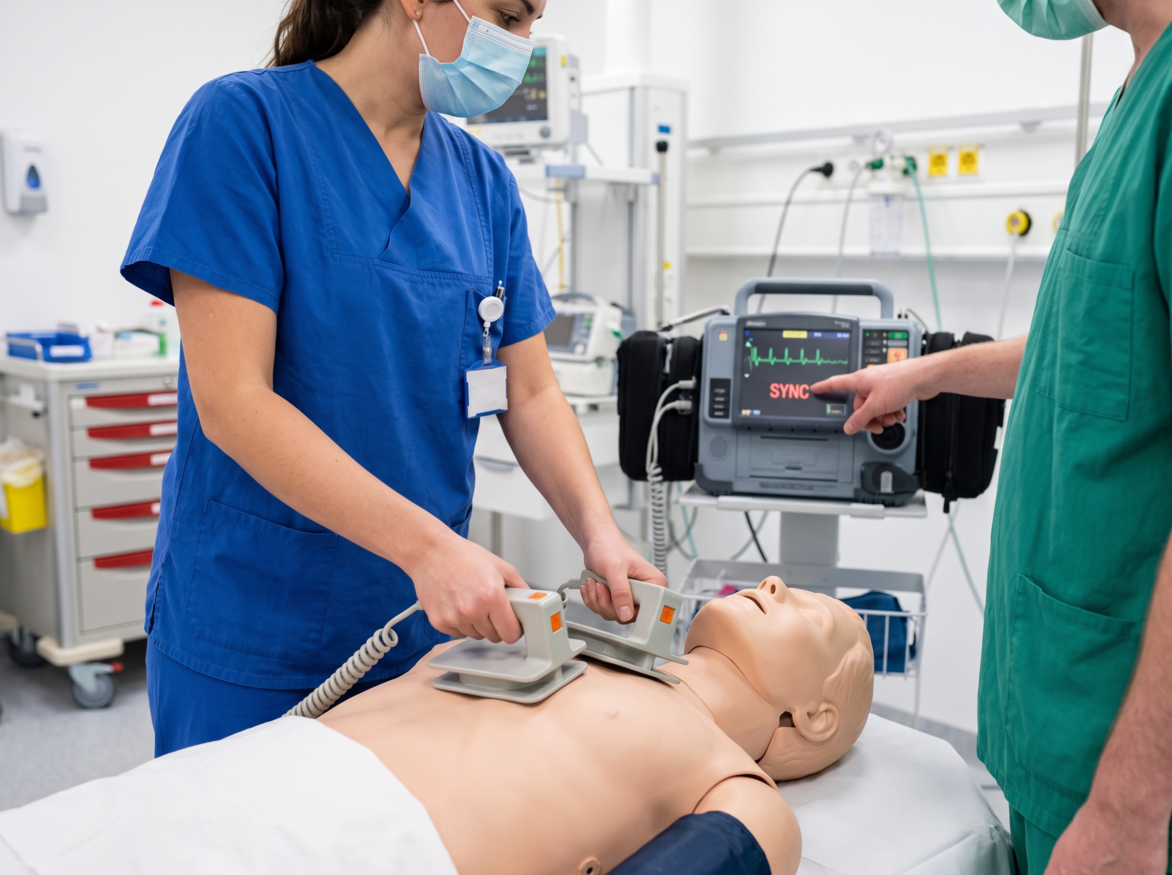
Healthcare providers preparing to deliver synchronized cardioversion during a simulated unstable tachycardia scenario


The Stable Branch: Four Boxes, Four Different Plans

If the patient is stable, the algorithm shifts from a hemodynamic question to an electrocardiographic one. You now sort the rhythm along two axes: is the QRS complex narrow or wide, and is the rhythm regular or irregular. Those two questions create four boxes, and each box points toward a meaningfully different management path. This is where the tachycardia algorithm earns its reputation as one of the more cognitively demanding sections of the ACLS course, because it asks you to hold two independent classifications in mind at once rather than pattern-matching to a single rhythm name.


It helps to picture the four boxes as a simple grid before working through each one individually.


  • Narrow and regular - usually SVT; vagal maneuvers and adenosine are the stable-patient first-line tools
  • Narrow and irregular - usually atrial fibrillation or a similar irregular supraventricular rhythm; rate control is the initial focus
  • Wide and regular - treat as ventricular tachycardia until proven otherwise; antiarrhythmic infusion is the stable-patient option
  • Wide and irregular - the highest-stakes box; several look-alike causes need different, sometimes opposite, treatments


None of these four labels are a final diagnosis by themselves. They are a starting orientation that tells you which family of treatments is even on the table before you narrow things down further with a 12-lead ECG, prior history, and how the patient responds to your first intervention.


Narrow and Regular

A narrow QRS complex with a regular rhythm most often represents a supraventricular tachycardia arising above or within the His bundle. The stable pathway here favors vagal maneuvers first, then adenosine if the rhythm persists and the maneuvers do not convert it. If the rhythm does not respond and remains stable, longer-acting agents such as a beta blocker or calcium channel blocker may be considered. Because this specific box has enough depth - dosing, repeat dosing, and what to do when adenosine does not work - to justify its own full write-up, the mechanics of vagal maneuvers and adenosine administration are covered in detail elsewhere; the point to hold onto here is simply that narrow-regular is the box where those tools apply.


Narrow and Irregular

A narrow QRS with an irregular rhythm points most commonly toward atrial fibrillation, though multifocal atrial tachycardia and atrial flutter with variable block can produce a similar irregular, narrow appearance. Rate control is typically the initial stable-patient strategy, often with a beta blocker or calcium channel blocker, while the underlying cause and duration of the arrhythmia are assessed. Anticoagulation status and the duration of the arrhythmia both factor into whether rhythm control is appropriate at this visit or should wait. The causes and treatment nuances of narrow-complex irregular tachycardia are worth a closer read, since this box has more clinical texture than a single-line algorithm box can capture.


Wide and Regular

A wide QRS complex with a regular rhythm is where the algorithm asks you to make a judgment call under real time pressure: is this ventricular tachycardia, or is it a supraventricular rhythm with aberrant conduction? The safe default in a stable patient is to treat a regular wide-complex tachycardia as ventricular in origin until proven otherwise, since VT is both more dangerous and more common in this presentation than aberrantly conducted SVT. Antiarrhythmic infusions such as procainamide, amiodarone, or sotalol are the stable-patient options, chosen based on the patient's other conditions. Adenosine may be considered only if the rhythm is regular and monomorphic, and only as a diagnostic and therapeutic trial, never as a substitute for treating a rhythm you suspect is VT with the respect it deserves.


Wide and Irregular

A wide, irregular rhythm is the box that should make you slow down rather than speed up. It can represent atrial fibrillation with aberrant conduction, atrial fibrillation with pre-excitation through an accessory pathway, or polymorphic ventricular tachycardia such as torsades de pointes. These have different treatments, and some of the drugs that are appropriate for one are actively dangerous for another - AV nodal blocking agents, for instance, can accelerate conduction down an accessory pathway in pre-excited atrial fibrillation and precipitate ventricular fibrillation. Expert consultation is appropriate here whenever it is available. For a deeper look at how wide-complex presentations get misread under pressure, the common myths around wide-complex tachycardia are worth reviewing before your next megacode or recertification attempt.


Where This Fits in the Bigger Algorithm Picture

The tachycardia algorithm does not exist in isolation. It shares a border with the bradycardia algorithm on the slow-rate side and with the cardiac arrest algorithm on the pulseless side - if a patient in front of you loses a pulse at any point during a tachycardia work-up, you stop following this algorithm and move immediately to the cardiac arrest algorithm instead. Keeping the boundaries between these algorithms clear is part of what the ACLS Algorithms Hub is designed to help with: seeing each algorithm not as a standalone flowchart to memorize but as one piece of a connected decision system that covers the full range of what a code team might encounter. Providers who study the tachycardia, bradycardia, and cardiac arrest algorithms together, rather than in isolation, tend to make faster and more confident branch decisions during an actual code, since they recognize the moment a patient crosses from one algorithm into another.


Using This Companion for the Megacode and Recertification

On a certification exam or in a megacode station, the tachycardia algorithm is usually tested by giving you a rhythm strip and a vital sign set, then asking what you do next. The fastest way to answer correctly is to run the same two questions every time, in order: first, does this patient meet criteria for instability, and second, if not, which of the four boxes does the QRS width and regularity put this rhythm into. Candidates who skip straight to naming the rhythm often talk themselves into the wrong branch, because rhythm recognition alone does not tell you whether cardioversion or medication is the right first move.


Pairing this companion with a rhythm-recognition resource makes the drilling faster. The ACLS rhythm recognition cheat sheet is built for exactly this kind of rapid-fire practice - look at a strip, classify it by width and regularity, then work through the same branching logic covered here until the sequence becomes automatic rather than something you have to reconstruct from memory under pressure.


Staying Sharp Between Codes

The tachycardia algorithm rewards providers who treat it as a decision framework rather than a chart to memorize once and forget. Reviewing the stability criteria, the reasoning behind treating the underlying cause versus the rate, and the four-box stable pathway on a regular basis keeps the logic accessible when a real tachyarrhythmia shows up on a busy shift rather than in a calm classroom setting. An ACLS certification or recertification course built around current AHA guidelines is a practical way to keep this algorithm - and the rest of the ACLS algorithm set - fresh, especially for providers who only run a handful of codes a year and want the decision tree to feel familiar rather than foreign when it matters. Whether you are certifying for the first time or coming back for recertification, working through the stability call, the unstable-to-cardioversion pathway, and the four stable boxes until they feel automatic is time well spent long before the next code ever starts.


ACLS Blogs

ACLS Tachycardia Algorithm Companion: Stable vs Unstable Decisions

The Tachycardia Algorithm Is a Decision Tree, Not a Rhythm List

Most providers picture the ACLS tachycardia algorithm as a rhythm strip quiz: identify the arrhythmia, then treat it. That is not how the algorithm is built. The Adult Tachycardia with a Pulse Algorithm is a branching decision tool, and every branch point is a question about the patient in front of you, not a question about the ECG in isolation. Get the branching logic right and the specific rhythm underneath becomes much easier to manage. Get it wrong, and you can shock a patient who did not need it or waste critical minutes on a stable-appearing patient who is actually crashing.


This companion walks through the algorithm the way it is actually built: first the stability call, then the fork between the unstable and stable branches, then the four boxes that stable tachycardia splits into based on QRS width and rhythm regularity. If you want the rhythm-by-rhythm mechanics of narrow-complex SVT specifically, including vagal maneuvers and adenosine dosing, that lives in its own decision card. Here, the goal is to make sure you never lose the thread of the algorithm's logic, even when the rhythm strip in front of you looks unfamiliar.

Emergency care team assessing a patient's cardiac monitor to judge hemodynamic stability during tachycardia


Step One: Is the Patient Stable or Unstable

Every pass through the tachycardia algorithm starts the same way: identify and treat the underlying cause, then assess whether the tachyarrhythmia itself is causing hemodynamic compromise. The algorithm defines instability using a specific, limited list of findings. A patient is considered unstable when the tachycardia is producing one or more of the following: hypotension, altered mental status, signs of shock, ischemic chest discomfort, or acute heart failure.


  • Hypotension - a blood pressure that is inadequate to perfuse the brain, kidneys, and myocardium
  • Altered mental status - new confusion, agitation, or decreased responsiveness that was not present before the rhythm change
  • Signs of shock - cool, pale, clammy skin, delayed capillary refill, or a weak thready pulse pointing to poor peripheral perfusion
  • Ischemic chest discomfort - chest pain or pressure consistent with myocardial ischemia that appeared or worsened with the fast rate
  • Acute heart failure - flash pulmonary edema, jugular venous distention, or new respiratory distress tied to the arrhythmia


Notice what is missing from that list: the heart rate itself. A number on the monitor is not, by itself, a criterion for instability. This is the single most common trap in tachycardia management, both in the resuscitation bay and on the certification exam. A young, otherwise healthy patient with a narrow-complex rhythm at 160 beats per minute who is talking to you, mentating normally, and maintaining a normal blood pressure is not unstable just because the number looks alarming. Conversely, a rate of 130 in a frail patient with new hypotension and confusion absolutely qualifies as unstable. The rate sets the stage; the clinical findings make the call.


Is the Tachycardia Causing the Instability, or Is Something Else Driving the Rate

The second trap sits right next to the first one, and it is the piece of reasoning that trips up exam candidates most often: before you treat the rate as the primary problem, ask whether the tachycardia is the cause of the instability or a compensatory response to something else. Sinus tachycardia from sepsis, hemorrhage, pain, fever, hypovolemia, or pulmonary embolism can produce a fast, sometimes irregular-looking rhythm alongside genuine hypotension or altered mental status. That patient is not a synchronized cardioversion candidate. Shocking sinus tachycardia does nothing for the underlying problem and can delay the fluids, blood, or source control the patient actually needs.


The distinction usually comes down to context and rhythm morphology rather than the rate alone. Sinus tachycardia rarely exceeds the physiologic maximum for age, tends to have an identifiable P wave before every QRS complex, and gradually speeds up or slows down rather than switching on abruptly. A primary tachyarrhythmia such as SVT, atrial fibrillation with rapid ventricular response, atrial flutter, or ventricular tachycardia is more likely to have an abrupt onset, a regular and unusually fast rate for a compensatory response, or a QRS morphology that does not fit a sinus mechanism. When the history and exam suggest an obvious driver such as fever, blood loss, or dehydration, treat that driver first and reassess the rhythm before reaching for the algorithm's unstable branch.


The Unstable Branch: Straight to Synchronized Cardioversion

Once you have confirmed that the tachyarrhythmia itself is the cause of the instability, the algorithm does not offer a menu of options. The unstable branch goes directly to synchronized cardioversion. There is no antiarrhythmic infusion to try first, no vagal maneuver to attempt, no waiting to see if the rate comes down on its own. Sedation should be given whenever the patient's condition allows, but the priority is restoring a stable rhythm before instability progresses to cardiac arrest.


A few operational details matter here regardless of the underlying rhythm. Synchronization mode must be confirmed and reconfirmed before each shock, since a defibrillator that loses sync on a subsequent shock can deliver an unsynchronized shock on the T wave. Pads or paddles should be placed to optimize current flow through the heart, and the team should be ready to move to immediate defibrillation if the patient deteriorates to a pulseless rhythm mid-procedure, since synchronized cardioversion has no role once the patient loses a pulse. For a narrow-complex regular rhythm like SVT, lower energy levels are typically effective; for atrial fibrillation, wide-complex tachycardia, or an irregular rhythm, higher starting energies are generally used. The exact energy selection is a rhythm-specific detail, but the branching decision - unstable means cardioversion, full stop - is the same regardless of what the underlying rhythm turns out to be.

Healthcare providers preparing to deliver synchronized cardioversion during a simulated unstable tachycardia scenario


The Stable Branch: Four Boxes, Four Different Plans

If the patient is stable, the algorithm shifts from a hemodynamic question to an electrocardiographic one. You now sort the rhythm along two axes: is the QRS complex narrow or wide, and is the rhythm regular or irregular. Those two questions create four boxes, and each box points toward a meaningfully different management path. This is where the tachycardia algorithm earns its reputation as one of the more cognitively demanding sections of the ACLS course, because it asks you to hold two independent classifications in mind at once rather than pattern-matching to a single rhythm name.


It helps to picture the four boxes as a simple grid before working through each one individually.


  • Narrow and regular - usually SVT; vagal maneuvers and adenosine are the stable-patient first-line tools
  • Narrow and irregular - usually atrial fibrillation or a similar irregular supraventricular rhythm; rate control is the initial focus
  • Wide and regular - treat as ventricular tachycardia until proven otherwise; antiarrhythmic infusion is the stable-patient option
  • Wide and irregular - the highest-stakes box; several look-alike causes need different, sometimes opposite, treatments


None of these four labels are a final diagnosis by themselves. They are a starting orientation that tells you which family of treatments is even on the table before you narrow things down further with a 12-lead ECG, prior history, and how the patient responds to your first intervention.


Narrow and Regular

A narrow QRS complex with a regular rhythm most often represents a supraventricular tachycardia arising above or within the His bundle. The stable pathway here favors vagal maneuvers first, then adenosine if the rhythm persists and the maneuvers do not convert it. If the rhythm does not respond and remains stable, longer-acting agents such as a beta blocker or calcium channel blocker may be considered. Because this specific box has enough depth - dosing, repeat dosing, and what to do when adenosine does not work - to justify its own full write-up, the mechanics of vagal maneuvers and adenosine administration are covered in detail elsewhere; the point to hold onto here is simply that narrow-regular is the box where those tools apply.


Narrow and Irregular

A narrow QRS with an irregular rhythm points most commonly toward atrial fibrillation, though multifocal atrial tachycardia and atrial flutter with variable block can produce a similar irregular, narrow appearance. Rate control is typically the initial stable-patient strategy, often with a beta blocker or calcium channel blocker, while the underlying cause and duration of the arrhythmia are assessed. Anticoagulation status and the duration of the arrhythmia both factor into whether rhythm control is appropriate at this visit or should wait. The causes and treatment nuances of narrow-complex irregular tachycardia are worth a closer read, since this box has more clinical texture than a single-line algorithm box can capture.


Wide and Regular

A wide QRS complex with a regular rhythm is where the algorithm asks you to make a judgment call under real time pressure: is this ventricular tachycardia, or is it a supraventricular rhythm with aberrant conduction? The safe default in a stable patient is to treat a regular wide-complex tachycardia as ventricular in origin until proven otherwise, since VT is both more dangerous and more common in this presentation than aberrantly conducted SVT. Antiarrhythmic infusions such as procainamide, amiodarone, or sotalol are the stable-patient options, chosen based on the patient's other conditions. Adenosine may be considered only if the rhythm is regular and monomorphic, and only as a diagnostic and therapeutic trial, never as a substitute for treating a rhythm you suspect is VT with the respect it deserves.


Wide and Irregular

A wide, irregular rhythm is the box that should make you slow down rather than speed up. It can represent atrial fibrillation with aberrant conduction, atrial fibrillation with pre-excitation through an accessory pathway, or polymorphic ventricular tachycardia such as torsades de pointes. These have different treatments, and some of the drugs that are appropriate for one are actively dangerous for another - AV nodal blocking agents, for instance, can accelerate conduction down an accessory pathway in pre-excited atrial fibrillation and precipitate ventricular fibrillation. Expert consultation is appropriate here whenever it is available. For a deeper look at how wide-complex presentations get misread under pressure, the common myths around wide-complex tachycardia are worth reviewing before your next megacode or recertification attempt.


Where This Fits in the Bigger Algorithm Picture

The tachycardia algorithm does not exist in isolation. It shares a border with the bradycardia algorithm on the slow-rate side and with the cardiac arrest algorithm on the pulseless side - if a patient in front of you loses a pulse at any point during a tachycardia work-up, you stop following this algorithm and move immediately to the cardiac arrest algorithm instead. Keeping the boundaries between these algorithms clear is part of what the ACLS Algorithms Hub is designed to help with: seeing each algorithm not as a standalone flowchart to memorize but as one piece of a connected decision system that covers the full range of what a code team might encounter. Providers who study the tachycardia, bradycardia, and cardiac arrest algorithms together, rather than in isolation, tend to make faster and more confident branch decisions during an actual code, since they recognize the moment a patient crosses from one algorithm into another.


Using This Companion for the Megacode and Recertification

On a certification exam or in a megacode station, the tachycardia algorithm is usually tested by giving you a rhythm strip and a vital sign set, then asking what you do next. The fastest way to answer correctly is to run the same two questions every time, in order: first, does this patient meet criteria for instability, and second, if not, which of the four boxes does the QRS width and regularity put this rhythm into. Candidates who skip straight to naming the rhythm often talk themselves into the wrong branch, because rhythm recognition alone does not tell you whether cardioversion or medication is the right first move.


Pairing this companion with a rhythm-recognition resource makes the drilling faster. The ACLS rhythm recognition cheat sheet is built for exactly this kind of rapid-fire practice - look at a strip, classify it by width and regularity, then work through the same branching logic covered here until the sequence becomes automatic rather than something you have to reconstruct from memory under pressure.


Staying Sharp Between Codes

The tachycardia algorithm rewards providers who treat it as a decision framework rather than a chart to memorize once and forget. Reviewing the stability criteria, the reasoning behind treating the underlying cause versus the rate, and the four-box stable pathway on a regular basis keeps the logic accessible when a real tachyarrhythmia shows up on a busy shift rather than in a calm classroom setting. An ACLS certification or recertification course built around current AHA guidelines is a practical way to keep this algorithm - and the rest of the ACLS algorithm set - fresh, especially for providers who only run a handful of codes a year and want the decision tree to feel familiar rather than foreign when it matters. Whether you are certifying for the first time or coming back for recertification, working through the stability call, the unstable-to-cardioversion pathway, and the four stable boxes until they feel automatic is time well spent long before the next code ever starts.


blogs

More articles for you

all articles

Start Today!

Get certified at your own pace, on your schedule, from the comfort of your home.